The more difficult question is whether the radiofrequency field reaches the anatomy evenly enough for that detail to remain trustworthy. In human tissue, the RF wavelength at ultra-high field becomes comparable to, or smaller than, the dimensions of the body. The resulting wave interference can produce regional signal loss, uneven flip angles, and contrast that changes across the field of view for reasons that have little to do with the underlying pathology.
This is the central problem behind parallel transmission in MRI and B1+ inhomogeneity. A 7T scanner may offer higher intrinsic signal-to-noise and new opportunities for structural, functional, and metabolic imaging, yet those advantages can be compromised when the transmit field is bright in one region and weak in another. Parallel transmission, or pTx, addresses the problem by replacing a single undifferentiated RF source with several independently controlled transmit channels, allowing the system to shape the electromagnetic field rather than accept its interference pattern as fixed.
The shift is technical, but its consequences are clinical. A more uniform excitation can mean fewer signal dropouts, more stable contrast, and a better chance that a subtle longitudinal change reflects biology rather than the position of a bright or dark region in the scanner.
The physics of B1+ inhomogeneity at 7 Tesla
MRI images are built from a sequence of carefully timed interactions between magnetic fields and tissue. The main static field, conventionally called B0, aligns nuclear spins. The RF transmit field, B1+, then applies the excitation that tips those spins away from alignment. The angle of that excitation—the flip angle—depends on the local amplitude and duration of the RF field.
At lower field strengths, the RF wavelength in tissue is generally large compared with the anatomy being imaged, so the transmit field can be treated as relatively smooth. At 7T, the situation changes. The wavelength becomes short enough for the RF waves emitted by different parts of the transmit system to interfere within the body. Depending on location, that interference may be constructive or destructive.
The resulting B1+ pattern is not a simple loss of brightness. It alters the way tissue is excited in the first place.
A region with a lower-than-intended B1+ amplitude may receive too small a flip angle. A region with excessive RF amplitude may be overexcited and may also absorb more energy than desired. The impact depends on the pulse sequence: a spoiled gradient echo, a spin echo, an inversion recovery acquisition, and an echo-planar functional scan will each respond differently to local variation in excitation.
In brain imaging, the consequences can appear in deep structures and in regions that are particularly sensitive to the geometry of the head and the placement of the RF coil. In body imaging, the problem can become even more pronounced because the anatomy is larger and tissue composition varies substantially across the field of view. At 7T, the liver, for example, presents a difficult environment for homogeneous excitation, and single-spoke static shimming may not be sufficient to prevent regional signal degradation.
At 7T, the transmit field is part of the image-forming problem. It cannot be treated as an invisible property of the scanner.
This matters because many quantitative and longitudinal methods assume that changes in signal can be interpreted biologically. If B1+ variation changes between subjects—or changes because the subject is positioned slightly differently—the measured difference may be partly technical. In a research protocol tracking cortical thickness, functional activation, perfusion, or metabolic signatures over time, that uncertainty can erode the value of an otherwise impressive acquisition.
The issue also reaches beyond image uniformity. RF energy deposited in tissue is described through specific absorption rate, or SAR. Ultra-high field MRI raises the importance of SAR management because the system must deliver RF energy at a higher frequency and because local peaks in absorption may not be visible from the reconstructed image alone. A transmit strategy that improves the flip angle while controlling local and global RF power is therefore doing two jobs at once: shaping contrast and managing safety constraints.
The hardware behind parallel transmission
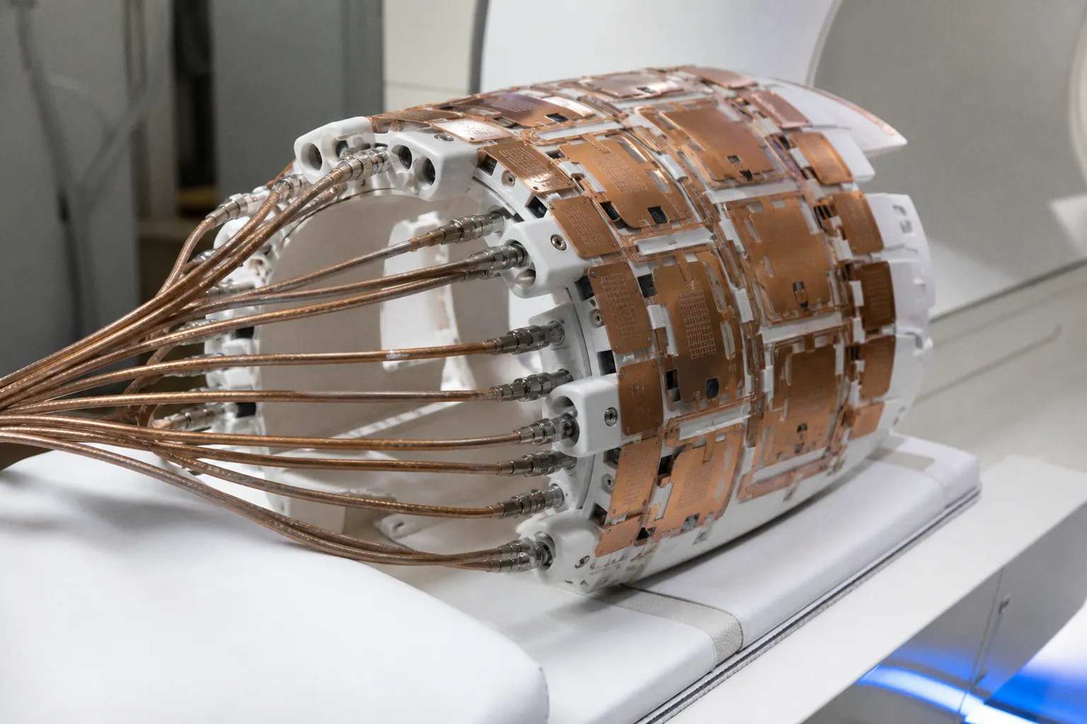
Parallel transmission begins with the transmit coil. Instead of using one effective transmit channel, a pTx system uses a local multi-channel transmit array, with elements that can be driven independently. Common configurations include 8, 16, or 32 transmit channels, although the practical arrangement depends on the scanner, coil design, anatomy, and intended application.
Each channel has its own RF waveform control and power-amplification pathway. The system can alter the amplitude and phase delivered through individual coil elements, creating a combined electromagnetic field that is more favorable for the anatomy being scanned.
This is not the same as simply increasing the number of receiver channels. Parallel reception and parallel transmission are related in the broader sense that both use spatially distributed coil elements, but their functions are different. Receiver arrays collect the signal after excitation. Transmit arrays determine how the tissue is excited before that signal is measured. A system may have sophisticated parallel reception without having the independent RF hardware required for dynamic pTx.
The transmit array is only one part of the architecture. A clinically usable system also requires:
- Independent RF waveform generation for each transmit channel, so that amplitude and phase can be controlled separately.
- RF power amplifiers capable of delivering the required energy to each element.
- A method for measuring or estimating the spatial distribution of B1+ across the subject.
- SAR monitoring and constraint management that can account for the combined contribution of all channels.
- Pulse-sequence software able to incorporate the calculated RF waveforms without disrupting timing, acceleration, or reconstruction.
- Safety interlocks and operational procedures appropriate to ultra-high-field imaging.
The relationship between hardware and software is unusually close here. A multi-channel coil without suitable pulse design may provide little more than a more complicated way of producing the same uneven field. Conversely, an elegant pTx pulse cannot be deployed safely if the scanner lacks the RF amplifiers, monitoring, and control pathways needed to deliver it.
The spatial arrangement of the array also matters. Coil elements positioned around the head or body produce fields with different local phases and amplitudes. pTx uses those differences as a form of control. By adjusting the contribution from each element, the system can reinforce excitation where the field is weak and reduce it where the field is excessive.
That correction is never completely abstract. It is tied to the anatomy, the loading created by the subject, and the pulse sequence itself. A configuration that works well for a particular brain acquisition may not transfer directly to abdominal imaging. Even within the same anatomical region, the best settings for a short gradient-echo scan may not be appropriate for a long spin-echo-based sequence.
Static B1 shimming: a useful first layer of correction
The simplest form of pTx control is static B1 shimming. The scanner selects a fixed phase and amplitude setting for each transmit channel, and those settings remain constant throughout the RF pulse. The goal is to create a more uniform combined B1+ field over the relevant anatomy.
Static shimming can be effective because it uses the spatial diversity of the transmit array without requiring the RF field to change during the pulse. It is comparatively straightforward to integrate into a workflow, and it can improve the distribution of excitation in regions where the primary problem is a stable spatial pattern of interference.
For some applications, that may be enough. The field can become more even, signal dropouts can be reduced, and the sequence may produce a more interpretable image without requiring a highly complex pulse design.
But static shimming has a fundamental limitation: a single set of amplitudes and phases must serve the entire RF pulse. The electromagnetic field produced by the body is not uniform, and the desired excitation may vary across space in a way that cannot be corrected by one fixed setting. Static B1 shimming therefore improves the field but does not make the underlying interference disappear.
This distinction becomes especially important in challenging body regions. In 7T liver imaging, multi-spoke pTx pulses have been shown to mitigate signal dropouts and provide better excitation homogeneity than single-spoke static B1 shimming. The additional control comes from changing the effective RF excitation over time rather than asking one fixed field configuration to solve every spatial problem at once.
A practical comparison looks like this:
| Feature | Static B1 shimming | Dynamic pTx pulse design |
|---|---|---|
| RF control | Fixed amplitude and phase settings during the RF pulse | Amplitude and phase can vary over time |
| Typical design logic | Find one field configuration that improves average uniformity | Build a time-varying excitation that addresses spatial variation throughout the pulse |
| Hardware burden | Requires multi-channel transmit hardware and control | Requires the same multi-channel platform plus time-resolved waveform delivery |
| Strength | Relatively direct implementation and lower sequence complexity | Greater freedom to correct severe or spatially complex B1+ patterns |
| Limitation | One fixed setting may not correct all regions or pulse demands | More demanding calibration, computation, safety control, and sequence integration |
| Representative approach | Single-spoke RF shimming | Multi-spoke pulses and other dynamic pTx designs |
The choice is not a contest in which dynamic pTx automatically replaces static shimming. In clinical research, reliability and workflow time matter alongside theoretical field uniformity. A simpler strategy that produces consistent images may be preferable to a more ambitious method that requires lengthy subject-specific preparation or is difficult to reproduce across scanner sessions.
Dynamic pTx and the logic of multi-spoke pulses
Dynamic pTx changes the RF shim as a function of time during the RF pulse. Rather than applying one spatially weighted excitation, the system delivers a sequence of RF subpulses—often described as spokes—separated by controlled changes in the excitation trajectory or the channel-specific RF settings.
Two-, three-, and four-spoke designs are examples of this approach. The number of spokes is not a universal measure of quality; it represents a design choice that must balance excitation performance, pulse duration, SAR, gradient demands, and the timing requirements of the sequence.
The conceptual advantage is that each spoke contributes a different spatial pattern. Together, they can approximate a target flip-angle distribution more effectively than one static configuration. Where one spoke produces a signal void, another may contribute a field that partially restores excitation. Where the combined RF power would otherwise concentrate locally, the pulse can be optimized to distribute the burden more carefully across channels and time.
This is why pTx pulse design is not merely an RF version of image post-processing. Once the image has been acquired with a severely underexcited region, software cannot reliably recreate the missing information. Dynamic pTx acts before signal collection, modifying the physical excitation so that the measured data begin from a more favorable state.
The approach has particular relevance to functional MRI. In GRE-EPI functional imaging, pTx spokes pulses have been associated with an improvement of up to 11% in temporal signal-to-noise ratio. That figure should not be interpreted as a universal gain for every subject, scanner, or protocol; temporal SNR is influenced by motion, physiological noise, reconstruction, sequence timing, and the stability of the experimental setup. Still, it illustrates the practical value of improving the excitation field in a sequence where susceptibility and signal instability can already make interpretation difficult.
Deep brain structures provide another example of why field control matters. Compared with a single-transmit arrangement, a pTx array has been reported to increase B1+ values by approximately 4.9% to 18.9% in structures including the caudate, insula, putamen, and temporal lobes. The effect is not identical across anatomy, and increased B1+ alone is not the same as improved diagnostic accuracy. The clinically relevant question is whether the revised field distribution produces more consistent contrast and more usable data for the sequence in question.
Consider the implications for studies of subtle degradation. A small regional difference in a single scan may be due to pathology, but it may also reflect B1+ variation, coil loading, or a change in head position. If the acquisition can reduce one source of spatial inconsistency, the remaining biological signal becomes easier to examine. This shift allows us to treat the scan less as a static picture and more as one observation along a trajectory.
Calibration, optimization, and time
Dynamic pTx usually depends on information about the subject-specific transmit field. Calibration measurements can estimate the B1+ pattern, after which an optimization routine determines the channel amplitudes and phases needed for the selected pulse. That preparation takes time, and the time cost can be clinically meaningful.
A research participant may tolerate a long protocol, but a patient undergoing a complex examination may have pain, cognitive impairment, involuntary movement, or limited ability to remain still. Every additional calibration step introduces another opportunity for motion or for the subject’s position to change. The estimated field may then no longer describe the anatomy with the same precision.
This is one reason universal pulses are attracting attention. They aim to provide a calibration-free pTx excitation that is robust across different subjects, using pulse designs derived to perform adequately over a population rather than optimized from scratch for every scan.
Calibration-free does not mean context-free. A universal pulse still depends on the hardware, the anatomy, the operating field strength, and the constraints imposed during design. It also represents a compromise: a pulse optimized for a broad range of subjects may not match the best possible subject-specific solution for any one individual. Its value lies in reducing preparation time while preserving a useful degree of B1+ homogenization.
For clinical workflows, that trade-off can be decisive. A method that is slightly less optimal in a simulation but consistently available at the scanner may have greater translational value than one that produces excellent results only after an elaborate calibration procedure.
SAR management is part of the image quality problem
At ultra-high field, the discussion of B1+ uniformity cannot be separated from RF energy deposition. The same independent control that allows pTx to reshape the field also creates a larger optimization problem: each transmit channel contributes to the total and local SAR pattern, and the combination can vary across space.
Global RF power is not the whole story. Local peaks may occur in specific anatomical regions, and a pulse that achieves excellent flip-angle uniformity may do so at an unacceptable SAR cost if the design is not constrained. For this reason, pTx optimization typically seeks a balance among several competing objectives:
- A target flip-angle distribution across the anatomy of interest.
- Reduced signal variation and fewer excitation dropouts.
- Acceptable global RF power deposition.
- Control of peak local SAR.
- A pulse duration compatible with the sequence.
- Gradient and hardware limits.
- Robustness to differences between the calibration model and the actual subject.
This balance explains why the visually most homogeneous solution is not always the operationally best one. A small residual B1+ variation may be acceptable if it allows a meaningful reduction in local SAR and produces a pulse that is more stable across subjects. In another sequence, preserving a specific contrast mechanism may justify a more sophisticated dynamic design.
The calculation of SAR is also a practical engineering concern. Commercial systems use vendor-specific methods and safety models, and the exact proprietary algorithms used for real-time pTx pulse recalculation are not generally transparent. That uncertainty should temper broad claims about what a given pTx implementation can guarantee. The hardware and pulse-design principles are shared, but the details of online monitoring, constraint handling, and sequence integration remain system-dependent.
Safety protocols therefore cannot be reduced to a software setting. Ultra-high-field imaging requires appropriate screening, validated coils and accessories, controlled operating procedures, and a clear understanding of how the selected pulse interacts with the scanner’s limits. Parallel transmission expands the available control space, but it also makes that space more consequential.
The best pTx pulse is not the one that makes the field look perfect in isolation; it is the one that preserves useful excitation, manageable SAR, and reproducible timing inside a real examination.
From better excitation to more reliable neuroimaging
The immediate output of pTx is a radiofrequency field. The reason to invest in that field, however, is not electromagnetic elegance. It is the quality of the biological inference that follows.
In neuroimaging, higher field strength can support finer spatial detail, stronger susceptibility contrast, and improved sensitivity for selected applications. Yet those gains are only helpful when the relationship between tissue and measured signal remains stable enough to interpret. B1+ inhomogeneity can introduce regional differences that complicate segmentation, quantitative mapping, functional activation analysis, and comparisons across participants.
A more homogeneous excitation can support several kinds of downstream work:
1. Structural imaging becomes easier to compare across regions.
If one part of the brain is consistently underexcited, tissue boundaries and regional contrast may become less dependable. pTx does not solve every source of bias, but it can reduce one major contributor to spatial variation.
2. Functional signals may become more stable.
EPI-based methods are sensitive to susceptibility, motion, physiological noise, and temporal drift. Better excitation can improve the usable signal before those other sources of variation are addressed.
3. Deep structures receive more balanced attention.
The caudate, putamen, insula, and temporal lobes are not interchangeable from an RF perspective. A transmit strategy that improves B1+ in these regions can make their measurements more consistent, although the effect must still be evaluated within the complete acquisition and analysis pipeline.
4. Longitudinal trajectories are easier to interpret.
In studies of neurodegeneration, treatment response, or cognitive reserve, the question is often whether a small change persists over time. Reducing avoidable technical variation strengthens the link between that measured change and the underlying biological trajectory.
5. Protocol design can become more ambitious.
When the transmit field is better controlled, researchers can explore sequences and contrasts that would otherwise be limited by signal dropout or inconsistent excitation. This does not mean that every 7T application becomes clinically ready, but it broadens the practical space for development.
The final point is important. pTx is an enabling technology, not a diagnostic conclusion. A more uniform B1+ field does not by itself prove that an acquisition is more sensitive to disease, that a biomarker will generalize across hospitals, or that 7T has replaced 3T for routine clinical imaging. Large, multi-center comparisons of diagnostic accuracy remain an important part of the evidence still needed.
Translational research must follow the whole chain: hardware, pulse design, safety constraints, reconstruction, image quality, reader performance, and patient outcome. An improvement at the RF stage is valuable, but its meaning depends on whether it survives the rest of that chain.
What universal pulses change in the clinical workflow
Subject-specific calibration is powerful because it responds to the individual anatomy in the scanner. Universal pulses are attractive because they respond to the constraints of clinical reality.
A calibration-free universal pulse can be prepared in advance and applied across a range of subjects, reducing the time required before imaging begins. This may be particularly useful when the protocol must be repeatable, when patient motion is a concern, or when a clinical service cannot accommodate a long optimization stage for every examination.
The phrase universal pulse should not suggest that one waveform is equally ideal for every patient. Human anatomy varies, and so do coil loading, tissue conductivity, head shape, body composition, and positioning. The goal is robust performance across that variation, not the elimination of variation itself.
That distinction becomes more important as pTx moves from research systems toward clinical environments. A method must be evaluated not only by its best-case B1+ map, but by the distribution of outcomes across ordinary subjects, imperfect positioning, interrupted calibrations, and routine operator decisions. It must also fit within the scanner’s existing scheduling and quality-assurance structure.
A useful clinical evaluation would therefore ask:
- Does the pulse reduce visible dropout in the anatomy relevant to the indication?
- Does it preserve or improve the contrast required by the sequence?
- How sensitive is performance to subject position and anatomy?
- What calibration or quality-control steps remain necessary?
- How does the method affect scan duration?
- Can SAR constraints be verified reliably during routine operation?
- Does improved field uniformity translate into better measurement repeatability or diagnostic confidence?
Recent developments, including clinical approval and comparative evaluation of pTx implementations on systems such as Siemens MAGNETOM platforms, indicate that the technology is moving beyond purely experimental demonstrations. Work on dynamic pTx SPACE sequences in brain MRI also reflects a broader effort to integrate time-varying transmit control into clinically recognizable sequence families. Even so, the path from technical approval to widespread clinical adoption is measured in reproducibility, training, maintenance, and evidence—not in the novelty of the pulse alone.
The next stage is integration, not spectacle
Parallel transmission makes 7T MRI more manageable by treating B1+ inhomogeneity as a controllable field problem. Multi-channel transmit coils provide the hardware degrees of freedom; static shimming offers a relatively direct correction; dynamic pTx pulses add temporal flexibility; universal pulses aim to reduce calibration burden; and SAR-aware optimization keeps those capabilities within a safe operating envelope.
The most important advance may be conceptual. Instead of accepting the RF field as a fixed limitation of ultra-high-field imaging, engineers and researchers can design the excitation around the anatomy, the sequence, and the clinical question. That design freedom is particularly valuable when the signal of interest is subtle and when studies depend on comparing people or tracking the same person across a long interval.
Still, the field should resist the temptation to turn improved B1+ uniformity into a promise of effortless clinical superiority. Parallel transmission does not erase motion, susceptibility, physiological noise, reconstruction bias, or the biological complexity of disease. It does not transform one scan into a complete account of a patient’s condition.
What it can do is quieter and, in practice, more consequential: make the excitation more even, the acquisition more reproducible, and the resulting measurements easier to connect to human biology. At 7T, that is not a minor engineering refinement. It is part of the groundwork required for high-field MRI to move from remarkable images toward dependable evidence.