The B1 field—the radiofrequency component that excites your protons—attenuates as it travels through tissue, and the further the anatomy sits from the receive structure, the more difficult it becomes to spend your SNR budget efficiently. For two decades, the workaround has been familiar: add receive channels, push parallel imaging harder, or, when finance allows, take the 7T leap whose capital cost, siting demands, and operating requirements make CFOs visibly flinch.
A growing body of peer-reviewed work now proposes a third route: engineered metamaterial structures that reshape the local RF field around the anatomy of interest, lifting SNR without raising the static magnetic field. The attraction is obvious. If the scanner cannot be made stronger, perhaps the field around the tissue can be made more useful. For anyone running a department, however, the question is not whether a resonant structure can produce a spectacular field map in a controlled setup. It is whether the gain survives real anatomy, real positioning, real reconstruction, and the unglamorous constraints of a clinical scanner.
The Physics of Localized B1 Field Manipulation: Beyond Static B0
Metamaterials in MRI are engineered passive electromagnetic structures composed of periodic arrays of sub-wavelength resonant elements. Depending on the design, those elements may be helical coils, split rings, capacitively loaded loops, coaxial resonators, or related structures tuned to interact with a particular band of the RF spectrum. For clinical scanners, that band sits at roughly 64 MHz at 1.5T and 128 MHz at 3T—the Larmor frequencies of hydrogen at those field strengths.
When an array is placed near the imaging volume, its resonators interact with the scanner's transmit field. The elements can store and re-radiate electromagnetic energy, changing the distribution of the local B1 field and concentrating it around the region where the anatomy lies. In practical terms, the metamaterial is not creating signal from nowhere. It is changing how the available RF energy is distributed in space.
That distinction matters because MRI signal is inseparable from geometry. A receive coil close to the anatomy generally sees a stronger local signal than one farther away, but a structure that produces a strong field at one location may not produce a useful field across the entire volume. The design target is therefore not simply maximum field amplitude. It is a workable compromise between enhancement, coverage, depth, loading, patient comfort, and compatibility with the rest of the coil assembly.
The crucial distinction is what these structures do not touch. They do not raise B0, the static field generated by the superconducting magnet. B0 is set by the scanner's magnet and operating conditions. What changes is the local B1 environment: the RF field used to excite the spins and the electromagnetic environment seen by the receive system.
By concentrating B1 where spins need to be excited and where the receive structure is most sensitive, a metamaterial can recover signal that would otherwise be lost to distance and tissue attenuation. By reshaping the B1 profile, it may also extend the useful coverage of a coil element or reduce a local field deficit. But the effect remains spatially conditional. A device optimized for a wrist, a superficial lesion, or a surface coil cannot automatically be treated as a general-purpose SNR layer for deep abdominal imaging.
The analogy to a directional antenna is useful, provided it is not taken too literally. A resonant structure can direct energy into a more useful region, much as an antenna can shape a radiofrequency beam. The MRI version operates at the Larmor frequency and inside a strongly loaded, lossy biological environment. Tissue conductivity, patient anatomy, coil loading, and the physical distance between the resonators and the target all influence the result. The body is not an empty test chamber, and the resonator does not behave identically once a patient replaces a phantom.
This is also why the 7T proposition has historically been described as “more magnet, more signal.” Increasing field strength changes the Larmor frequency and, in principle, provides access to more signal per unit time. It also brings its own problems: B1 nonuniformity, stronger susceptibility effects, more demanding RF management, and a different operational and capital-cost profile. Metamaterials attempt to capture part of the practical benefit without changing B0. They are not a substitute for 7T. They are a way of making a local portion of an existing RF system behave more efficiently.
The word local should stay in every serious discussion of metamaterials in MRI RF coils. It is the boundary between a useful engineering result and a misleading headline.
Quantifying SNR Gains: From Clinical Wrist Imaging to 32-Fold Boosts
The literature spans a wide range of SNR figures, and the spread tells its own story. Laboratory demonstrations, surface-coil experiments, and clinically oriented extremity studies are measuring related effects under very different geometries. The numbers should therefore be read as a map of design possibilities, not as a single performance curve.
| Reported result | Setup | Field strength | Geometry or context |
|---|---|---|---|
| 1.26×–1.40× SNR | Wireless metamaterial insert paired with a body coil versus a conventional transmit/receive extremity coil | 1.5T | Clinical wrist imaging |
| 33% wider sensitive field of view | Same wireless metamaterial extremity setup | 1.5T | Wrist imaging |
| Approximately 4.2× localized SNR | Array of magnetic metamaterial resonant elements | Scanner-class relationship not specified in the demonstration | Localized region near the array |
| 66% SNR improvement | Near-field coupling array, or NFCA, metasurface versus a wired coil with equivalent coverage | 3T-class | Surface imaging |
| More than 94% acceleration-factor retention | NFCA metasurface used with parallel imaging | 3T-class | Surface imaging |
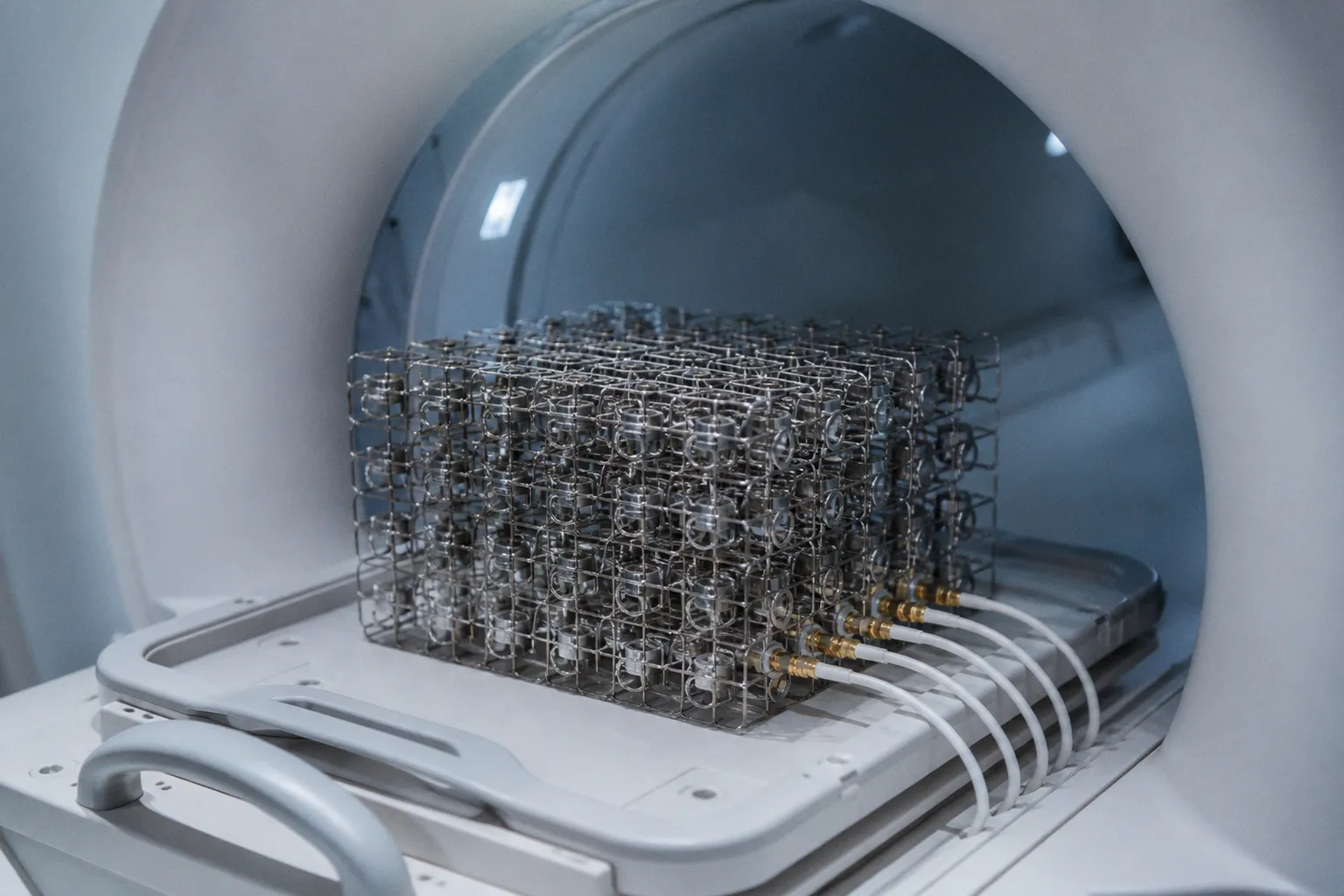
| Up to 32× | Circularly polarized coaxial-cable metamaterial cage operated with a body coil | 3.0T | Localized region near the cage structure |
Two patterns stand out.
First, the clinically oriented and surface-imaging results cluster much closer to the 1.3×–1.7× range than to the spectacular laboratory maximum. The wireless wrist result is important precisely because it addresses a recognizable clinical use case rather than only demonstrating that a resonant array can manipulate a field. The NFCA result is also notable because it combines a substantial reported SNR improvement with a parallel-imaging measurement.
Second, the 32× result is a localized, geometry-dependent measurement. It comes from a coaxial cage operated in close proximity to a body coil, where the metamaterial structure reshapes B1 over a small, defined region. That is a striking physics result. It is not a uniform upgrade to the entire field of view, and it does not mean a department can replace its phased-array infrastructure with a cage.
The 32-fold number is not a coil. It is a physics demonstration. Decide which one you are reading.
The same discipline is needed with the smaller numbers. A reported SNR ratio depends on what is being compared, where the measurement is taken, how the noise is estimated, and whether the result comes from a phantom, a volunteer, or a patient examination. It also matters whether the comparison is made at equal spatial resolution, equal scan time, equal acceleration, or equal image quality. A metamaterial may raise signal in a region while also changing noise correlation or the spatial sensitivity profile of the receive system. The number on the graph is not automatically the number that appears as a shorter protocol or a sharper diagnostic image.
For a scanner administrator, the 1.26×–1.66× results are the more relevant part of the current record because they sit closer to deployable surface and extremity applications. The approximately 4.2× result is a useful demonstration of localized B1 concentration and a design reference for future structures. The 32× result belongs at the top of the research leaderboard. It demonstrates what a carefully tuned geometry can do; it does not by itself establish a procurement case.
There is another practical distinction between SNR enhancement and clinical value. Extra signal can be spent in several ways:
- to reduce acquisition time while preserving image quality;
- to increase spatial resolution without accepting the same noise penalty;
- to support stronger fat suppression, diffusion weighting, or other demanding contrasts;
- to reduce the number of averages required by a protocol;
- or simply to produce a more robust examination when positioning and patient motion are not ideal.
Those are not interchangeable benefits. A department that already completes a wrist protocol comfortably may gain little from an accessory that only makes the same three-minute scan look better. A research protocol limited by low signal, resolution, or repeated acquisitions may value the same enhancement much more. The right question is not whether the metamaterial increases SNR. It is what the department can do with the additional SNR.
Architectural Innovations: Metasurface Inserts vs. Near-Field Coupling Arrays
Several structural approaches now dominate the experimental record. Each makes a different trade-off between local enhancement, depth penetration, mechanical packaging, and compatibility with existing coil hardware.
Passive metasurface inserts
A passive metasurface insert is usually a planar or gently curved array of resonant elements placed on or near the anatomy. Some designs operate wirelessly and are intended to sit between the patient and the scanner's existing RF hardware. Their role is to concentrate or redistribute B1 in the immediate vicinity of the structure.
In the 1.5T wrist work, this approach delivered 1.26×–1.40× SNR compared with a conventional transmit/receive extremity coil and produced a 33% wider sensitive field of view. The appeal is straightforward: the insert can function as an accessory rather than as a complete replacement for the scanner's body coil or the department's entire receive architecture.
That apparent simplicity hides the engineering problem. The insert must be positioned consistently, remain comfortable, avoid obstructing patient access, and behave predictably when loaded by different wrists and different body geometries. A wireless device also has to be designed around detuning, resonance stability, and safe operation under the scanner's RF transmit conditions. “Passive” does not mean operationally invisible.
The near-field nature of the effect also limits the depth at which the insert can remain useful. A device close to a wrist or a superficial target has a favorable geometry. Move the target farther away and the enhancement generally becomes less pronounced. This is why a strong extremity result should not be generalized to deep pelvic, abdominal, or thoracic imaging without direct evidence.
Active hybrid receive arrays with integrated resonators
A second approach integrates metamaterial elements into a conventional receive array. Instead of placing a separate insert over the anatomy, designers embed capacitively loaded ring resonators or similar structures into the two-dimensional layout of the coil.
The attraction is control. An integrated design can be tuned as a single system, allowing the engineer to balance peak surface SNR against penetration depth, element spacing, coupling, and mechanical conformity. It may also provide a more stable relationship between the resonant structures and the receive channels than a loose accessory placed on top of an existing coil.
The price is complexity. Once the resonators are part of the receive array, they affect fabrication, tuning, decoupling, servicing, and the way the coil behaves under different loading conditions. The design no longer has the simplicity of an independent passive layer. It becomes a new coil architecture, even if it retains the external shape and workflow of a familiar one.
This hybrid family is therefore where much of the vendor-facing engineering interest lies. It offers a route to practical packaging, but it also creates more points at which integration can fail. The relevant question is no longer just whether a resonator can enhance B1. It is whether the complete array can be tuned, characterized, cleaned, positioned, repaired, and supported like equipment that belongs in a clinical inventory.
Near-field coupling array metasurfaces
The near-field coupling array, or NFCA, represents a different connection strategy. Instead of relying on a conventional wired, or galvanic, connection between the metasurface and the receive chain, the architecture couples through the inductive near field.
The reported 2025 NFCA work found a 66% SNR improvement relative to a wired coil with equivalent coverage. More importantly for workflow, the same architecture was tested with parallel imaging and retained more than 94% of the acceleration factor of the comparison coil. That result is specific to the NFCA architecture and its reported experiment. It should not be transferred automatically to every passive insert or hybrid array.
The attraction of the NFCA design is that it addresses two problems at once: local field enhancement and a connection architecture that may preserve useful channel behavior. But “may” is doing real work here. Parallel-imaging performance depends on the spatial sensitivity profiles of the receive channels, not simply on the amount of signal visible in a single image. A metasurface that raises local signal while making channel profiles less distinct can create a poor geometry factor and give back the gain during accelerated reconstruction.
Coaxial metamaterial cages
A more exotic architecture is the circularly polarized coaxial-cable metamaterial cage associated with the 32× demonstration at 3T. The cage is a useful research platform because it can create a strong, controlled resonant environment around a defined region. It is also the least natural fit for a routine clinical accessory.
Cages are bulkier, less anatomically conformal, and harder to package around variable patient anatomy. They raise practical questions about access, positioning, patient comfort, cleaning, emergency removal, and compatibility with existing coils. The fact that a structure can produce a large local enhancement does not tell us whether it can be deployed in a high-throughput imaging room.
The shared design thread across these approaches is that the metamaterial acts as a passive electromagnetic lens, refocusing or redistributing B1 where the designer wants it while the scanner continues to provide transmit power, signal digitization, and image reconstruction. But that does not make the accessory “RF-transparent” in every operational sense. The scanner may not need a new pulse sequence, yet the system still has to be characterized for loading, coupling, detuning, transmit behavior, and safety. Hybrid coils may require additional vendor integration, and the boundaries of responsibility between the device and the scanner remain unresolved.
Hybrid Integration and Parallel Imaging Compatibility
The single biggest workflow threat from any new coil technology is breaking parallel imaging. A radiologist who gains 1.4× SNR but loses a substantial amount of acceleration has not necessarily improved the examination. The extra signal may simply be converted into longer acquisitions, longer breath-holds, or lower throughput. That is why the acceleration-factor retention reported for the NFCA work is more operationally meaningful than its SNR headline alone.
Parallel imaging depends on the spatial sensitivity profiles of individual receive channels being sufficiently distinct to reconstruct undersampled k-space. The system is using those differences as information. A metamaterial that uniformly amplifies signal across an array may improve a conventional image while contributing little to accelerated reconstruction. A design that changes the profiles in an uncontrolled way may do worse: it can increase local signal and still impose a reconstruction penalty.
The NFCA result is encouraging because it reports both enhancement and retention of parallel-imaging performance. It suggests that near-field coupling can be engineered without automatically destroying the channel diversity required for acceleration. That is a meaningful result, but it remains an architecture-specific result. The current evidence does not justify saying that wireless wrist inserts, NFCA metasurfaces, and integrated hybrid arrays all preserve comparable acceleration behavior.
If a coil upgrade breaks your parallel-imaging factor, you have bought SNR with scan time. That is not an upgrade.
The integration problem becomes harder when passive inserts are stacked over existing phased arrays. In principle, the insert can enhance the local RF environment without replacing the receive channels. In practice, the additional resonant layer can alter coupling, loading, and the spatial response of the underlying coil. Positioning becomes part of the experiment. A few millimeters, a change in orientation, or a different patient contour may change the balance between enhancement and unwanted interaction.
More aggressive designs integrate resonators directly into the coil layout. That can make the system more stable and easier to characterize as a single device, but it also pushes more responsibility into the coil design. Engineers have to manage resonance, element-to-element coupling, cable behavior, preamplifier interaction, and the effects of patient loading. The scanner's reconstruction software may remain unchanged, but the hardware feeding that reconstruction is no longer conventional.
The same is true on the transmit side. A passive structure can interact with the transmit field even when it has no active electronics. The local electric and magnetic fields may change, and the scanner's existing assumptions about RF deposition may not fully describe the new arrangement. SAR validation is necessary, but it is not a complete description of system integration. Depending on the architecture, vendor collaboration may be needed to establish operating limits, document the intended coil configuration, and determine how the device is represented in the scanner's safety and service workflows.
This distinction matters to hospital IT and engineering teams. An accessory that requires no new pulse sequence may still require a new acceptance procedure. The site may need a defined placement protocol, a device-specific safety review, quality-control testing, and a clear answer to what happens when the scanner is serviced or upgraded. If the vendor does not formally support the device, the burden does not disappear; it moves to the institution.
There is also a reconstruction question. Higher raw SNR does not guarantee higher diagnostic utility if noise becomes spatially correlated or if the enhancement is confined to a portion of the image. A useful evaluation should examine uniformity, artifact behavior, channel covariance, accelerated acquisitions, and the stability of the result across loading conditions. The most persuasive demonstration is not a single high-SNR image. It is a protocol-level comparison showing what the department can gain without creating a new failure mode.
That is where the difference between a research insert and a clinical coil becomes visible. A research setup can optimize positioning for each experiment. A clinical device must tolerate the ordinary variability of a busy department: different technologists, different patients, different body habitus, and different levels of patience at the scanner table.
Current Limitations and the Path to Clinical Standardization
Three constraints still separate the laboratory numbers from the procurement conversation, with a fourth running through all of them: the evidence base is not yet broad enough to treat one successful anatomy as a general platform.
Depth penetration
Metamaterial enhancement is fundamentally a near-field effect. The closer the resonant structure sits to the target anatomy, the more pronounced the local field manipulation is likely to be. The 1.26×–1.66× results reported for surface and extremity imaging fit that physical picture. They involve anatomies where the device can be placed close to the region of interest and where the geometry is relatively controlled.
Deep anatomy is a different problem. In pelvic, abdominal, and thoracic imaging, the relevant tissue may sit farther from any practical insert, and the surrounding anatomy may load the resonant structure in less predictable ways. Published results show the expected dependence on depth: enhancement falls as the target moves away from the structure. That does not make deep-imaging applications impossible, but it means they require their own designs and their own validation.
A wrist insert should therefore be judged as a wrist insert. It may be an excellent solution for extremity imaging without offering a meaningful lift to a prostate or liver protocol. The broader the anatomy, the harder it becomes to preserve a strong local effect while maintaining useful coverage and avoiding regional intensity variation.
Patient loading and reproducibility
A resonant element is tuned to an electromagnetic environment, and the patient is part of that environment. Tissue conductivity, body habitus, positioning, and nearby conductive materials can shift the behavior of the structure. A device that performs well with one loading condition may require retuning or careful tolerance design to behave consistently across patients.
The issue is not limited to image quality. Loading changes can affect the local RF field and safety assessment. Conductive implants and other hardware may introduce additional interactions. Motion can change the relationship between the resonator and the anatomy. A device intended for routine use has to be robust to these variations rather than merely impressive under ideal placement.
This is one reason clinical extremity work is valuable. The wrist is not trivial, but it is a relatively shallow and controlled target. Results there help establish whether a concept can survive patient use. They do not close the question for every other anatomical region.
Product, regulatory, and vendor status
The product and regulatory path remains unresolved. It is not safe to state that every architecture has failed to complete a particular vendor-neutral approval route, because the public record does not establish a settled status across all relevant designs. At the same time, the available research does not justify treating metamaterial accessories as standardized, plug-and-play products for arbitrary Siemens, GE, or Philips systems.
The uncertainty is practical as much as bureaucratic. RF inserts interact with the scanner's transmit field and with assumptions embedded in the vendor's validated SAR model. A hybrid receive array may require its own tuning, interface, calibration, and service documentation. A passive insert may still need device-specific testing and a defined operating configuration. Regulatory clearance, vendor acceptance, research-use deployment, and routine clinical availability are different milestones. They should not be collapsed into one claim about whether the technology is “approved” or “not approved.”
For an academic site, an unresolved path may be manageable through a research collaboration, institutional review, and engineering support. For a community hospital, the same uncertainty can stop a purchase before the SNR discussion begins. Procurement teams need a supported device, a defined scanner compatibility list, a safety file, training requirements, and a service model. The current metamaterial literature does not yet provide those things uniformly across architectures.
System integration beyond SAR
SAR validation is essential, but it is not the only system-side issue. A new resonant structure can affect transmit-field distribution, coil loading, detuning behavior, receive-channel coupling, calibration, and the conditions under which the scanner's existing monitoring remains valid. Whether the scanner needs software changes depends on the architecture and the vendor's integration model; it should not be assumed either way.
A hybrid coil may behave differently from a passive overlay because the resonators are part of the active receive assembly. An NFCA design may require a different characterization approach from a wired coil. A cage that works with a body coil may have very different mechanical and operational requirements from a wrist insert. Each configuration needs its own evidence.
The right standard is therefore not “the scanner does not know the metamaterial is there.” The right standard is that the scanner, coil, accessory, and site workflow have been characterized well enough that the device can be used within documented limits. Until that evidence exists, the technology remains promising but configuration-dependent.
What standardization would have to demonstrate
A clinically credible metamaterial platform would need more than a high local SNR ratio. It would need to show, across a defined scanner and anatomy range, that:
- the enhancement is reproducible across patients and positioning conditions;
- the device does not introduce unacceptable artifacts or intensity nonuniformity;
- parallel-imaging behavior is measured for the specific architecture rather than inferred from another design;
- transmit-field changes and SAR behavior are understood for the intended operating configuration;
- the device can be cleaned, positioned, removed, and serviced within ordinary clinical workflows;
- the claimed gain translates into a useful change in scan time, resolution, contrast, or robustness;
- and the vendor, regulator, and hospital engineering team agree on who owns integration and ongoing support.
That is a demanding list because MRI accessories are not evaluated in isolation. They become part of a coupled system. A resonator, receive array, scanner transmit chain, reconstruction pipeline, patient, and technologist all contribute to the final result.
Final Position
Metamaterials in MRI RF coils are not a substitute for higher B0 or for simply adding more receive channels. Anyone who sells them that way is selling brochure optimism, and the reading room has had enough of that. What they offer is a genuine, physics-grounded lever for lifting local SNR, particularly in surface and extremity imaging where the resonant structure can sit close to the target anatomy.
The current evidence supports a more measured conclusion. Gains in the 1.3×–1.7× range are the most relevant to near-term clinical discussion because they appear in clinically oriented and surface-imaging configurations. The approximately 4.2× result shows how powerful localized B1 concentration can be under a controlled design. The 32× result demonstrates the upper edge of what a carefully arranged coaxial structure can produce in a defined region. None of those numbers should be read as a universal multiplier for every protocol.
The NFCA work is particularly important because it reports parallel-imaging retention alongside SNR improvement. That makes it more than a signal-amplitude story. But the finding belongs to the NFCA architecture and its tested configuration. It does not establish that every wireless insert or integrated hybrid array will preserve the same acceleration behavior.
For departments evaluating this technology, the practical decision is to connect the physics to an actual bottleneck. If the problem is shallow anatomy, limited surface sensitivity, or a protocol where additional signal can be exchanged for resolution or acquisition time, a metamaterial accessory may become a legitimate upgrade path. If the problem is deep-tissue coverage, inconsistent positioning, or an unsupported scanner configuration, a spectacular local field map will not solve it.
The evidence should be demanded at the level of the proposed use: the same anatomy, the same scanner class, the same coil arrangement, and the same reconstruction strategy. The device should come with a clear account of safety validation, loading behavior, integration requirements, and the limits of any parallel-imaging claim. Product availability, regulatory status, and vendor support should be treated as open implementation questions until they are documented for the specific system.
That leaves metamaterials in an interesting but narrow position. They are no longer just an elegant electromagnetic trick, but they are not yet a universal clinical platform either. For surface and extremity work, the technology has crossed into serious engineering territory. For deep anatomy and broad departmental deployment, the decisive work is still ahead: reproducibility, system integration, clinical validation, and standardization. Until those pieces are in place, the strongest metamaterial result belongs in the research discussion—not automatically in the capital plan.