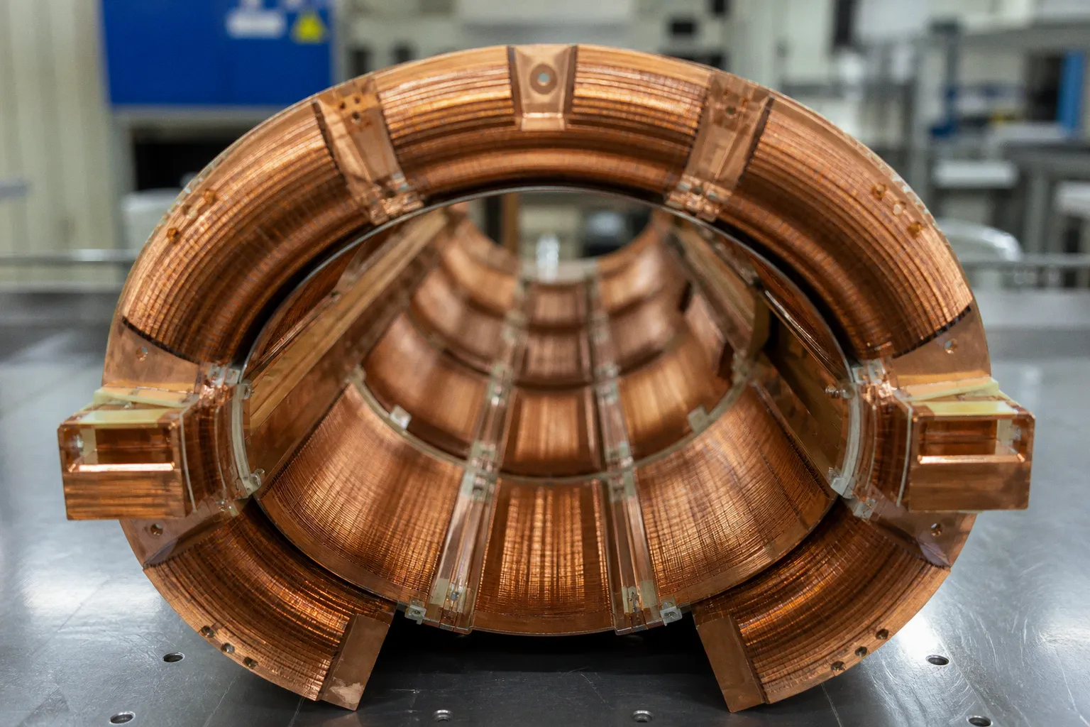
Philips’ BlueSeal 1.5T magnets, for example, use a factory-sealed load of approximately 7 liters—about 0.5% of the helium volume associated with a conventional 1.5T system.
That is not a cosmetic reduction. It changes the engineering boundary of the scanner. The magnet becomes lighter. Installation loses one major piece of infrastructure. Routine helium refilling is removed. The quench-management problem is reduced, although not erased.
The harder question is not whether sealed systems consume less helium. They do. The harder question is whether helium-free MRI scanner technology can preserve the field stability, uptime, safety behavior, and protocol envelope demanded by high-field clinical imaging. The answer is not universal. It depends on what “helium-free” means, which field strength is involved, and whether the comparison is being made at the magnet level or at the level of diagnostic performance.
The physics of cryogenics: from open bath to sealed micro-cooling
Superconducting MRI magnets require their coil assemblies to remain near absolute zero. Traditional systems use liquid helium with a boiling point near −269°C. The superconducting wire operates inside a cryogenic environment that provides the thermal margin required to maintain zero-resistance behavior under the magnet’s static field and operating conditions.
The conventional architecture is an open helium bath. The magnet coils are immersed in a large reservoir. This provides substantial thermal mass, but it also creates a permanent logistical dependency. The helium inventory is large. The cryostat is heavy. The installation requires dedicated quench-vent infrastructure to route rapidly expanding helium gas away from occupied spaces if the magnet undergoes a quench.
A sealed system takes a different path. The cryogenic load is reduced sharply, and the cooling system is designed to maintain the superconducting magnet without regular replenishment from an external helium supply. In the BlueSeal architecture, the liquid helium is factory sealed. The system still contains helium. “Helium-free” is therefore an operational description, not a literal statement that the magnet contains no helium at all.
This distinction matters. A sealed 7-liter load is not equivalent to zero cryogen. It is, however, a fundamentally different maintenance model from an open-bath magnet holding approximately 1,500 liters.
The thermal problem is moved from bulk cryogen storage toward controlled conduction and cryocooler-based heat removal. The superconducting coils are cooled through a thermal pathway rather than relying on direct immersion in a large open reservoir. That pathway must tolerate the heat generated by the cryocooler, environmental fluctuations, mechanical vibration, and the transient thermal behavior of the magnet during operation.
The magnet still has to do the same mathematical job. It must create a highly uniform static field. It must remain stable over the acquisition window. It must preserve the field conditions assumed by shimming, frequency calibration, fat suppression, spectroscopy, diffusion imaging, and other sequence-specific operations.
“Helium-free” does not mean cryogen-free. It means that the cryogenic burden has been compressed, sealed, and engineered into the magnet rather than managed as a recurring facility supply.
Helium-free MRI versus traditional helium cooling
The relevant comparison is not simply reservoir volume. It is a stack of coupled engineering constraints.
| Parameter | Traditional open-bath MRI | Sealed or micro-helium MRI |
|---|---|---|
| Cryogen inventory | Approximately 1,500 liters of liquid helium in a conventional superconducting system | Single-digit liters in current sealed designs; BlueSeal 1.5T systems use about 7 liters |
| Helium replenishment | Historically dependent on external helium supply and service logistics | Factory-sealed load designed to eliminate routine refilling |
| Quench infrastructure | Traditional systems require dedicated quench venting | Fully sealed systems can eliminate traditional quench pipes |
| Magnet mass | Higher, because of the large cryostat and helium reservoir | Lower for certain sealed designs; reductions of up to 2,050 kg have been reported for some magnets |
| Installation flexibility | Constrained by venting, structural load, and room design | Better suited to elevated floors, constrained sites, and mobile installations |
| Field-strength maturity | Broad installed base across 1.5T and 3.0T clinical workflows | Strongest operational history in lower-field sealed systems; 3.0T equivalence remains an active question |
| Main technical risk | Helium supply, quench management, cryostat mass | Cooling architecture, thermal stability, serviceability, and protocol validation |
The table exposes the central issue. Sealed cooling removes infrastructure constraints, but it does not eliminate the need for magnet engineering. It relocates the difficulty.
The scanner is not only a magnet
A magnet can be easier to install and still produce a clinically inferior examination if the rest of the acquisition chain is poorly matched to its field environment. MRI performance is produced by the interaction of several systems:
- The static field \(B_0\) determines the nominal Larmor frequency, susceptibility behavior, chemical-shift separation, and part of the available signal.
- The gradient coils determine spatial encoding, diffusion encoding, echo-planar trajectory, slew-rate limits, and acoustic behavior.
- The radiofrequency chain determines transmit efficiency, receive sensitivity, specific absorption rate constraints, and parallel-imaging performance.
- The pulse sequence determines how those physical limits become an image, spectrum, or quantitative map.
- The reconstruction determines how undersampling, motion, noise, and trajectory errors are converted into usable or misleading output.
A sealed magnet changes the first layer. It does not automatically improve the gradient system, RF coil design, k-space trajectory, or reconstruction algorithm.
This is where comparisons between helium-free MRI and traditional helium cooling often become imprecise. The cooling architecture is visible. The image-quality consequences are distributed across the system and may not be attributable to cooling alone.
A 1.5T sealed scanner with mature gradients, a properly tuned RF chain, and validated reconstruction can be a highly competent clinical instrument. A conventional open-bath 1.5T system with degraded gradients, aging coils, or unstable service support can perform worse in practice. Conversely, a high-field sealed system must demonstrate more than a stable magnet. It must preserve the protocol-specific behavior expected by radiologists and researchers.
The physical metrics remain the same:
- signal-to-noise ratio, or SNR, under defined acquisition conditions;
- geometric distortion in echo-planar imaging;
- temporal stability across repeated acquisitions;
- field homogeneity after shimming;
- gradient fidelity and eddy-current behavior;
- RF transmit uniformity and SAR behavior;
- susceptibility sensitivity at the target anatomy;
- reconstruction robustness under motion and undersampling.
A magnet cooling label does not substitute for these measurements.
Infrastructure evolution: removing the quench pipe
The clearest practical advantage of sealed systems is architectural. Traditional MRI installations must account for a quench. If superconductivity is lost rapidly, stored magnetic energy is released and liquid helium can transition into gas. The expansion is extreme. A quench pipe is designed to direct that gas outside the building rather than into the scan room.
Removing the traditional vent path has consequences beyond construction cost. It changes where the scanner can be placed. Elevated floors, compact urban medical sites, mobile trucks, and rooms with limited external routing become easier to evaluate. The design burden shifts toward the magnet’s internal containment and cooling architecture.
This is not a license to treat quench risk as irrelevant. The static magnetic field remains dangerous. Ferromagnetic objects can still become projectiles. Patient screening, access control, emergency procedures, implant verification, and zone management remain fundamental MRI safety requirements.
The sealed architecture also does not make the room indifferent to failure. A facility still needs documented procedures for magnet faults, power loss, cryocooler failure, service access, and controlled shutdown. It needs to understand whether a specific scanner is fully sealed, micro-helium, or merely low-consumption. These are not interchangeable categories.
Weight is another meaningful constraint. Some sealed BlueSeal magnet configurations are reported to reduce weight by up to 2,050 kg compared with conventional systems. That difference can affect structural reinforcement, transportation, crane requirements, and installation sequencing. It can also reduce the number of sites rejected before procurement begins.
But weight reduction has to be read against the complete system. The scanner is not just the magnet. Patient table, gradients, RF cabinets, shielding, cooling hardware, power electronics, and room construction still contribute to the installed load. A lighter magnet improves the boundary condition. It does not turn a high-field MRI suite into ordinary building equipment.
Global helium dependency is an imaging problem
MRI is not the only consumer of helium, but it is a major one. Medical MRI accounts for approximately 32% of global helium consumption according to the supplied industry reporting. That percentage gives the engineering shift its strategic importance.
Open-bath systems expose hospitals to a supply-chain dependency that is easy to underestimate during normal operation. Helium is not produced as a primary commodity in the same way as many industrial gases. Availability depends on extraction, purification, transport, storage, and regional market conditions. The scanner may be clinically essential, but its cryogen supply is connected to a wider industrial system.
The risk is not limited to the price of a refill. A delayed delivery can extend downtime. A service intervention may require specialized logistics. A quench can convert a clinical event into a facility event, with consequences for scheduling, patient access, and capital planning.
Sealed magnets attack that dependency at the source. The scanner is charged once during manufacture and designed to operate without routine liquid-helium replenishment. This reduces the recurring inventory requirement and makes the system less sensitive to local helium logistics.
The environmental argument is therefore strongest at the infrastructure level:
1. Less helium is stored at each installation.
2. Routine refill operations are removed.
3. Transport and handling requirements are reduced.
4. Quench vent construction may be avoided.
5. Magnet mass can be reduced in certain configurations.
6. Mobile and constrained-site deployment becomes more plausible.
None of this proves that a sealed magnet has a lower total environmental burden in every lifecycle analysis. Manufacturing the conduction-cooling assembly, operating cryocoolers, servicing electronics, and eventually decommissioning the scanner all consume energy and materials. The correct comparison requires a lifecycle model, not a single helium-volume figure.
Still, the helium reduction is not marginal. Moving from approximately 1,500 liters to 7 liters changes the operational profile by orders of magnitude. It constrains the facility less. It does not eliminate physics.
From 1.5T foundations to 3.0T systems
The 1.5T environment is where sealed-helium technology has established its clearest foundation. Philips introduced BlueSeal operations in 2018, and the company reported its 1,111th BlueSeal installation in May 2024. Those milestones indicate substantial deployment, but they do not by themselves establish universal equivalence across vendors, field strengths, or clinical protocols.
The 3.0T problem is more demanding.
At 3.0T, the RF frequency is higher. Susceptibility effects are more pronounced. B0 and B1 inhomogeneity become more disruptive in anatomically difficult regions. SAR constraints can restrict sequence timing and flip-angle choices. Echo-planar imaging becomes more sensitive to field imperfections and distortion. Spectroscopy and quantitative imaging demand stable frequency behavior and reproducible shimming.
A 3.0T magnet therefore has to preserve more than nominal field strength. It must maintain a usable operating envelope for the sequences that justify 3.0T in the first place.
At RSNA 2025, Philips introduced the BlueSeal Horizon platform, described as a 3.0T MRI system using a helium-free magnet design. That is an important hardware milestone. It is not the final clinical verdict. Robust multicenter evidence across advanced protocols remains an area of ongoing study.
The validation burden should be protocol-specific. A scanner may match expectations for routine brain imaging while requiring more careful characterization for:
- high-resolution susceptibility-weighted imaging;
- diffusion and distortion-sensitive tractography;
- functional MRI with long echo trains;
- magnetic resonance spectroscopy;
- quantitative relaxation mapping;
- high-resolution musculoskeletal imaging;
- abdominal imaging where respiratory motion and B0 variation interact;
- multi-site research protocols that depend on harmonized signal behavior.
The correct question is not whether a helium-free 3.0T magnet “works.” Almost any modern system can produce images. The question is whether it yields stable, comparable, and diagnostically acceptable data under the exact protocol burden imposed by the site.
The engineering transition is already credible at the installation layer. The unresolved question is protocol-level equivalence, especially where 3.0T exposes every weakness in homogeneity, RF control, and reconstruction.
Zero boil-off MRI is not the same category
The term “zero boil-off” creates another source of confusion. A zero-boil-off system is designed to recondense helium gas and minimize net helium loss during operation. It can dramatically reduce consumption compared with older open-bath systems. But it still relies on a substantial cryogenic architecture and does not necessarily provide the same installation profile as a sealed micro-helium magnet.
A sealed magnet and a zero-boil-off magnet solve related problems through different engineering strategies.
| Architecture | Primary objective | Helium behavior | Facility implication |
|---|---|---|---|
| Open-bath superconducting magnet | Maintain coils in a large liquid-helium reservoir | Large stored volume; refill and quench logistics remain central | Requires conventional cryogenic planning and quench venting |
| Zero-boil-off MRI | Recondense helium and minimize routine loss | Very low net consumption, but substantial cryogenic infrastructure remains | Reduces refill dependence without necessarily removing installation constraints |
| Sealed micro-helium magnet | Operate with a small factory-sealed helium load | Single-digit-liter inventory in current examples | Can remove routine refilling and traditional quench pipes |
| Fully helium-free concept | Remove helium from the operating magnet architecture | Depends on the underlying cooling and superconducting technology | Requires separate validation; should not be conflated with sealed-helium designs |
For procurement and technical review, the terminology should be made explicit. Ask whether the system contains sealed liquid helium. Ask whether the magnet is conduction cooled. Ask whether the quench pipe is genuinely unnecessary under the manufacturer’s installation specification. Ask what service event occurs if the cryocooler fails. Ask whether the claimed helium-free designation describes the magnet, the whole scanner, or only the absence of routine refill.
A precise label prevents an imprecise comparison.
What clinical and research teams should measure
A replacement decision should begin with the protocol library, not the marketing category. The scanner must be evaluated against the work it is expected to perform.
For routine 1.5T neuroimaging, the relevant question may be whether sealed cooling preserves expected SNR, motion tolerance, scan time, and uptime while reducing site complexity. For advanced neuroimaging, the test becomes more severe. Researchers may need temporal stability, cross-session reproducibility, phase consistency, distortion characterization, and compatibility with existing preprocessing pipelines.
The acquisition audit should include:
- Static-field homogeneity. Measure residual B0 variation after shimming across the anatomy and volume of interest. A nominal field value is insufficient.
- SNR and g-factor behavior. Compare identical coils, acceleration factors, voxel sizes, and reconstruction settings where possible. Otherwise, the comparison is contaminated by receive-chain differences.
- Gradient performance. Review maximum amplitude, slew-rate limits, duty-cycle behavior, eddy-current correction, and actual sequence timing. Nominal gradient figures do not fully describe trajectory fidelity.
- EPI distortion. Characterize geometric displacement and susceptibility-driven signal loss in diffusion and functional protocols.
- RF uniformity and SAR. At 3.0T, B1 variation and SAR constraints can alter sequence behavior even when the images appear acceptable at a glance.
- Thermal and temporal stability. Repeated acquisitions should test drift, frequency consistency, and calibration behavior over the operating day.
- Motion correction interaction. A reconstruction can suppress visible motion while changing quantitative signal properties. The pipeline must be validated, not merely inspected.
- Service continuity. Record the failure modes of the cryocooler, power electronics, and control systems. The absence of helium refills does not imply the absence of downtime.
A research site also needs data harmonization plans. If a multicenter study moves from open-bath to sealed magnets, sequence parameters may remain nominally identical while the effective signal behavior changes. The study should treat the scanner transition as a hardware covariate. Phantom data, repeat scans, and site-specific calibration become more important, not less.
The practical balance: where sealed systems already win
The strongest case for sealed-helium MRI is not speculative. It is operational.
A site with difficult structural access can benefit from lower magnet mass. A mobile MRI platform can benefit from the removal of a traditional quench pipe. A hospital exposed to helium supply interruptions can reduce one recurring vulnerability. A facility planning a new installation can gain room-layout flexibility.
These benefits are especially tangible when the alternative is not a new open-bath system but a constrained renovation. In that context, the magnet’s infrastructure requirements can determine whether deployment is feasible at all.
The economics are more conditional. Lower helium consumption does not automatically mean lower total cost. Cryocoolers, compressors, maintenance contracts, replacement parts, electrical demand, and service response all affect the lifecycle balance. A procurement model that counts only helium savings is incomplete.
The clinical argument is also conditional. At 1.5T, sealed designs have a meaningful installed base and a credible operational record. At 3.0T, the technology is advancing, but the evidence must be read protocol by protocol. The missing piece is not a vague sense of promise. It is independent, multicenter equivalence across the full range of high-field applications.
The verdict
Helium-free MRI is ready to replace traditional liquid-helium architecture in many installation scenarios. It is not yet a universal replacement for every superconducting MRI workflow.
The distinction is technical. Sealed micro-helium magnets reduce cryogen inventory from approximately 1,500 liters to single-digit liters. They can eliminate routine refilling. They can remove traditional quench pipes. They can reduce magnet weight by up to 2,050 kg in certain configurations. Those are established engineering advantages.
They do not, by themselves, prove identical performance across every diffusion, spectroscopy, functional, quantitative, or high-resolution 3.0T protocol. That claim requires measured SNR, field stability, gradient fidelity, RF behavior, distortion control, and longitudinal reproducibility.
The transition is therefore not from “helium” to “no helium.” It is from bulk cryogen dependence to tightly engineered cooling. The facility burden falls. The validation burden remains.
A sealed magnet is ready when the site values installation flexibility and operational resilience, and when the protocol evidence is specific enough to support the intended clinical or research workload. Anything broader is not a conclusion. It is a label.