The Twenty-Nine Percent Problem
In a study of adult surgical candidates whose 3T MRI scans were negative, parallel transmit (pTx) 7T MRI revealed previously unseen structural lesions in 9 of 31 patients — roughly 29% of the cohort. The same study found that imaging findings altered clinical management in 18 of 31 cases, or about 58%.
Those are not incremental numbers. They do not prove that every newly identified lesion led to a resection plan, and they do not tell us that all of these patients had previously been sent back to medical therapy. They establish something more useful and more defensible: in a group selected precisely because conventional imaging had not provided a structural explanation, pTx-enabled 7T changed what clinicians knew and, in many cases, changed what they did next.
That distinction matters. The clinical value of an advanced scanner is not measured only by whether it produces a sharper image. It is measured by whether the image changes the diagnostic conversation, the next test, the multidisciplinary review, or the treatment pathway.
And yet, for most of the past decade, the 7T scanner parked in a hospital’s research wing has been exactly that: a research-wing artifact. A physics marvel with a clinical workflow problem the size of the temporal lobe. The first 7T MRI system received FDA clearance for clinical diagnostic imaging in October 2017, when the Siemens MAGNETOM Terra became the first ultra-high-field scanner approved for routine diagnostic use. The bet was that higher field strength would translate directly into better diagnostic yield.
The bet was right in principle and incomplete in practice. The problem was never simply the magnet. The problem was the RF field.
The magnet was the easy part. The RF field has been the bottleneck, and pTx gives clinicians a way to manage the most stubborn part of that problem.
The Physics of the 7T Barrier: Why B1+ Inhomogeneity Stalled Clinical Adoption
To understand why 7T MRI spent so long as a research tool, you have to understand what happens to radiofrequency fields at ultra-high field strengths. At 7 Tesla, the effective wavelength of the RF pulse in tissue becomes short relative to the dimensions of the head. That creates dielectric shading, standing-wave effects, and pronounced B1+ transmit-field inhomogeneity.
B1+ is the component of the transmit RF field that drives the excitation of the tissue. In a conventional single-transmit system, the scanner has limited control over how that field is distributed across the patient’s anatomy. At 3T, the resulting variation is usually manageable for routine brain imaging. At 7T, it can become the defining limitation of the examination.
The practical consequence is uneven excitation. One region of the brain may receive an effective flip angle while another receives too little. Signal intensity can vary across the head, with the anterior temporal lobes, brainstem, and cerebellum particularly vulnerable to loss of uniformity in some protocols and patient anatomies. The result is a peculiar failure mode: exquisite detail in one part of the brain and inadequate diagnostic confidence in another.
For a patient with drug-resistant focal epilepsy, that is not a cosmetic flaw. The regions affected by transmit-field variation can overlap with the regions where a subtle cortical malformation, hippocampal abnormality, or other structural correlate may matter most. A higher nominal field strength does not automatically produce a clinically useful image if the excitation is poorly distributed.
This is the B1+ barrier. It is not a software bug, and it is not something that can be removed by simply increasing the field strength again. It is a fundamental constraint of ultra-high-field RF transmission. For years, 7T protocols were therefore compromises: strong signal and high spatial resolution where the field behaved well, less reliable contrast and coverage where it did not.
That is why the early clinical story of 7T was so uneven. The scanner could reveal structures that were difficult to resolve at lower field strengths, but the benefit depended heavily on anatomy, sequence design, patient positioning, calibration, and the local RF environment. A research protocol could be optimized around those constraints. A clinical service has to deliver a reproducible examination across a much wider range of patients.
There are other physical problems as well. B0 inhomogeneity can distort frequency-sensitive sequences, susceptibility effects become more pronounced, and motion remains motion. pTx does not erase those limitations. It addresses a specific and important part of the 7T problem: the distribution of the transmit field and the way RF power is managed during the examination.
The 8-Channel Breakthrough: How pTx Architecture Reshapes RF Transmission
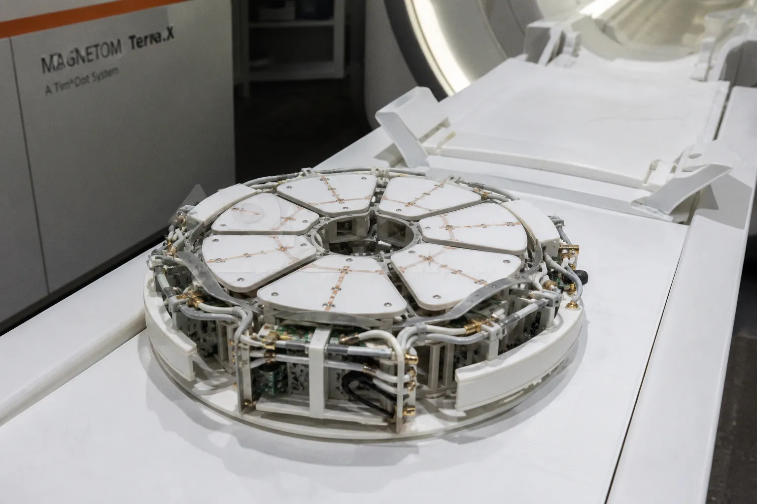
Parallel transmit is the architectural answer to the B1+ problem. Instead of one large RF transmission path exciting the entire anatomy with a single amplitude and phase relationship, pTx uses an array of independent transmit channels. Each channel can be controlled separately, allowing the system to adjust the relative amplitude and phase of the RF waveform across the head.
The eight-channel system that received FDA clearance in March 2024 made that approach more visible as a clinical product rather than a research capability. The clearance of the Siemens MAGNETOM Terra.X marked an important step for an 8-channel pTx architecture in clinical 7T MRI. Auburn University installed the first field-installed, clinically approved Terra.X that same month, another practical milestone for a technology that had spent years being demonstrated in research environments.
The key phrase is RF shimming. By changing the contribution of individual transmit channels, the system can shape the composite B1+ field to reduce regional dropouts and improve the consistency of excitation. It is not magic, and it is not identical to correcting every voxel independently in real time. The scanner is using calibrated hardware, patient-specific or protocol-specific adjustments, and carefully designed pulse sequences to produce a more usable field distribution.
That distinction is important because pTx is often described as if it solves “the physics” of 7T. It does not. It mitigates a major RF transmission problem. It can also help manage SAR, which becomes a central constraint as field strength rises, but it does not make motion irrelevant, eliminate B0 artifacts, or guarantee that every sequence will be robust for every patient.
In the reading room, however, the intended benefit is straightforward. Better transmit-field control can improve visualization in regions that are vulnerable to signal loss. The brainstem and cerebellum may become more consistently evaluable. Temporal-lobe imaging can become less dependent on a favorable combination of anatomy and positioning. The improvement is not merely a matter of making an already adequate image prettier; in selected cases, it can determine whether the examination answers the clinical question at all.
The operational contrast looks like this:
| Operational factor | Single-transmit (CP) 7T | 8-channel parallel transmit (pTx) 7T |
|---|---|---|
| B1+ uniformity | More vulnerable to regional shading and signal variation | More control over the spatial distribution of the transmit field |
| Temporal-lobe imaging | Can be limited by local signal loss | Better suited to mitigating transmit-field dropouts |
| Brainstem and cerebellar coverage | May be inconsistent in affected protocols | Greater potential for usable signal across the posterior fossa |
| SAR management | Can constrain flip angle, repetition time, or sequence design | More flexible RF power distribution, within system and protocol limits |
| Sequence development | Often dependent on research optimization | Requires validated clinical protocols and ongoing local maintenance |
| Reproducibility | Sensitive to anatomy, positioning, and calibration | Improved control, but still dependent on setup and patient-specific factors |
That last row is the one that matters to the MR technologist at 4:30 on a Friday. A sequence that works beautifully in a carefully selected research cohort is not automatically a sequence that works when the schedule is full, the patient is anxious, and the technologist has to repeat the examination tomorrow if the most important series fails.
Repeat acquisitions are the silent throughput killer. Every dropped sequence in the temporal lobe becomes a possible callback, a rescheduled patient, a difficult explanation to the referring clinician, and another delay before the case reaches a presurgical conference. pTx can reduce one source of failure, but it does not remove the operational cost of motion, coil positioning, incomplete screening, or a protocol that has not been tuned for the institution’s patient population.
Clinical Impact in Epilepsy: Translating High-Field Imaging into Surgical Decisions
The 31-patient epilepsy study is, for now, one of the clearest pieces of clinical evidence that pTx-enabled 7T MRI can affect management in a difficult population. The cohort consisted of adult surgical candidates with negative 3T MRI examinations — patients for whom standard imaging had not identified a structural lesion despite a clinical reason to keep looking.
This is the right population for testing the technology. A study of unselected patients with obvious lesions would tell us that 7T produces high-resolution images. A study of 3T-negative candidates asks the harder question: can ultra-high-field imaging reveal information that is not already available, and is that information clinically consequential?
In 9 of 31 patients, pTx 7T revealed a structural lesion that had not been seen previously. In 18 of 31, the imaging findings were significant enough to alter clinical management. The facts support those two findings. They do not support the stronger claim that all nine patients received a surgical roadmap, a defined resection plan, or a direct route to the operating room. Imaging can change management in several ways: it can direct additional electrophysiological evaluation, reshape the differential diagnosis, change the interpretation of prior studies, influence eligibility for a procedure, or support a decision to continue investigating.
That is still a meaningful result. For a 3T-negative epilepsy patient, the discovery of a previously unseen lesion can change the center of gravity of the case. The team may have a new anatomical hypothesis to test against semiology, EEG, PET, SPECT, or invasive monitoring. A lesion may become a target for closer review even when it does not by itself settle the question of resection.
The comparison between transmission modes adds another layer. Lesions were better visualized with pTx than with single-transmit CP mode in 57% of cases, and CP mode was not judged better for lesion visualization in the study. That does not mean pTx wins every sequence or that the result can be generalized to every scanner and every protocol. It does show that the hardware configuration mattered within the examined workflow.
The direction of the finding is important. pTx was not simply being used to produce a marginally cleaner version of the same scan. It changed the conditions under which high-field signal was delivered to the brain. The diagnostic advantage was therefore tied to the transmission architecture, not only to the number printed on the magnet.
In epilepsy imaging, the useful question is not whether 7T looks sharper. It is whether the additional signal reveals an abnormality that changes the next clinical decision.
There is also a temptation to turn the 9-of-31 result into a promise about surgical outcomes. That would be a mistake. Finding a lesion is not the same as proving that it causes the seizures. A lesion that is visible at 7T still has to be reconciled with the patient’s history, seizure semiology, scalp EEG, functional mapping, and, where appropriate, invasive recordings. The scan can create a better hypothesis. It cannot replace the rest of the presurgical process.
This is where responsible reporting of advanced MRI matters. The value of pTx is already substantial without claiming that every newly visible lesion produces a defined operation. Eighteen altered management decisions in a carefully selected 31-patient cohort is a clinical signal. It is not a guarantee of treatment, and it is not yet a population-wide estimate of how often routine 7T will change care.
Workflow Integration and the Path to Sub-20-Minute Acquisition Protocols
Here is where pragmatic skepticism earns its keep. The clinical evidence is real. The regulatory clearance is real. What remains uncertain is whether 7T with pTx can fit into a hospital workflow built around scheduling, staffing, safety, and repeatability rather than around the best possible research image.
The sub-20-minute acquisition target for accelerated high-resolution brain and knee clinical examinations is the benchmark toward which vendor development has been moving. The 0.8 mm isotropic 3D voxel resolution used for T1, T2, FLAIR, and EDGE sequences at 7T is competitive with high-resolution 3T examinations in the right protocol, but the comparison is conditional. Acceleration has to work as designed, and a fast acquisition is useful only if it remains diagnostically reliable.
Motion correction, parallel imaging, and compressed sensing each come with sequence-specific trade-offs. They need to be tuned to the anatomy, contrast, acceleration factor, and reconstruction pipeline. A protocol that is efficient for a cooperative adult undergoing a structural brain examination may behave differently in a patient with frequent movements, anxiety, pain, or difficulty following instructions.
The shorter the advertised examination, the more important the hidden preparation becomes. Patient screening has to account for the 7T environment. Positioning and coil setup need to be consistent. Prescan calibration must be reliable. The technologist has to understand when a field-map or calibration result is good enough, when a sequence needs to be repeated, and when the problem is likely to be motion rather than RF inhomogeneity.
That is the difference between acquisition time and room time. A scan may contain fewer minutes of actual sequence execution while still requiring more preparation, troubleshooting, or review than a familiar 3T protocol. A hospital evaluating clinical 7T adoption should therefore measure the whole pathway: patient arrival, screening, setup, calibration, acquisition, reconstruction, radiologist review, and recovery of failed or incomplete series.
Staffing is part of the physics story because the scanner does not operate independently of the people around it. A 7T system is not a 3T scanner with a larger magnet. Siting requirements, RF shielding, cryogen considerations, SAR monitoring, implant screening, and emergency procedures all become more demanding. The staff must also recognize that pTx introduces additional calibration and quality-control responsibilities. More transmit channels create more control, but they also create more system behavior to monitor.
The peripheral device ecosystem is another practical constraint. The October 2024 FDA clearance of Bayer’s MEDRAD MRXperion injection system for use at field strengths up to 7T was a small but telling milestone. It suggests that the supporting equipment needed for routine 7T examinations is beginning to catch up with the scanner hardware. A clinical service depends on that ecosystem: injectors, monitoring equipment, coils, patient accessories, safety procedures, and software all have to be compatible with the intended use.
For a hospital considering a clinical 7T program, the relevant questions are operational rather than promotional:
- Can the MR physics team maintain pTx calibration, protocol performance, and quality assurance over time?
- Do technologists have enough training to manage the longer setup and the additional safety and RF considerations?
- Are the radiologists prepared to interpret ultra-high-field examinations with sequence contrasts and artifacts that may differ from their 3T experience?
- Can the institution define which referrals genuinely benefit from 7T rather than sending every difficult case into a more complex scanner?
- Is there a process for correlating new findings with EEG, prior imaging, and multidisciplinary review?
- Can the service absorb occasional extended examinations when motion, breath-hold failure, or a calibration problem disrupts the planned protocol?
The answer to the last question matters more than the brochure version of throughput. A fast protocol that fails unpredictably can be less useful than a slightly longer protocol that produces dependable diagnostic data. Clinical adoption is not achieved by reaching a target acquisition time once. It is achieved when the protocol performs consistently enough to be scheduled, staffed, interpreted, and defended as part of patient care.
Regulatory Milestones and the Future of Routine Ultra-High-Field Diagnostics
The regulatory arc is worth tracing because it shows how the technology has moved from a scientific demonstration toward a clinical platform.
October 2017 brought the first 7T MRI clinical clearance in the United States. March 2024 brought clearance for an 8-channel pTx architecture on the MAGNETOM Terra.X. October 2024 brought FDA clearance of a peripheral injection system for use at field strengths up to 7T. Each step is incremental, but the cumulative effect is important: the scanner is no longer the only regulated component being considered in isolation. The broader clinical environment is beginning to support ultra-high-field imaging.
That does not mean the regulatory path is complete. The status of specific clinical pTx sequences and applications varies by jurisdiction, and the installed base of clinical 7T systems remains small compared with 1.5T and 3T. Claims about “routine” adoption should therefore be treated with the same skepticism working radiologists apply to any vendor roadmap. A cleared system can be used clinically; it does not automatically create a mature service line.
The more realistic question is where 7T adds enough value to justify its complexity. In the near term, the strongest case remains specialized imaging: complex neurological cases, selected epilepsy evaluations, high-resolution structural examinations, and musculoskeletal applications where additional field strength and tailored RF transmission can answer a question left unresolved at lower field.
That is a meaningful clinical niche, but it is not the same as a wholesale replacement for 3T. The relative role of each field strength will depend on local referral patterns, equipment availability, patient mix, staffing, protocol maturity, and reimbursement. Future improvements could expand the role of 7T in some hospital networks. At the same time, 3T will remain the more flexible backbone for broad routine imaging in many settings because it is more widely deployed and supported by established workflows.
The division of labor is therefore likely to be more useful than the replacement narrative. A 3T scanner can handle high-volume general neuroimaging and provide the familiar platform for most referrals. A pTx-enabled 7T system can serve as an escalation tool when the clinical question justifies higher spatial resolution, greater susceptibility sensitivity, or a second look at anatomy that remains unexplained.
Regulatory clearance opens the door to clinical use. It does not, by itself, build the workflow on the other side of the door.
The next phase will be less about proving that pTx can improve a field map and more about proving that institutions can reproduce the clinical benefit. That requires larger cohorts, standardized protocols, transparent reporting of failed and incomplete examinations, and follow-up that distinguishes a newly visible lesion from a lesion that genuinely changes treatment. It also requires outcome studies that avoid collapsing every management change into a surgical success.
For vendors, the challenge is to make the system easier to operate without hiding the complexity that still matters. For hospitals, the challenge is to build a service around patient selection and clinical coordination rather than around the prestige of owning a 7T magnet. For radiologists and physicists, the task is to define where pTx makes a measurable difference and where a well-executed 3T study remains the better choice.
The Verdict
Parallel transmit 7T MRI has crossed an important line. It is no longer accurate to describe the technology only as a research curiosity. The 8-channel architecture cleared in 2024, the early epilepsy evidence, and the developing peripheral-device ecosystem all point toward a genuine clinical role.
But a clinical role is not the same as a routine role. The 9-of-31 lesion finding and the 18-of-31 management result show why the technology matters: in selected 3T-negative epilepsy patients, pTx-enabled 7T produced information that was not available from the prior examination and changed clinical management in a substantial portion of the cohort. They do not establish that every newly seen lesion creates a surgical plan, and they do not justify treating the scanner as a shortcut around the presurgical process.
The physics is not “solved.” pTx mitigates B1+ transmit-field inhomogeneity and provides more control over RF power and SAR. That is a major advance, particularly for anatomy that has historically suffered from signal loss at 7T. Motion, B0 inhomogeneity, susceptibility, patient tolerance, calibration, and sequence validation remain part of the real examination.
The workflow is equally unfinished. A hospital buying a pTx-equipped 7T scanner is buying a specialized imaging capability, not a guaranteed throughput upgrade. The institution also needs trained technologists, MR physicists, validated protocols, compatible peripheral equipment, careful patient selection, and a clinical team able to act on the findings.
Nine out of thirty-one is a real number. So is eighteen out of thirty-one. Together, they make the strongest case for 7T pTx today: not that it will replace 3T across the hospital, but that it can become a high-value escalation tool when conventional MRI has not answered the question.
The research tool is becoming a clinical instrument. The harder work now is making the instrument dependable enough to belong in everyday care.