It can also be wrong in exactly the regions where anatomy changes position, shape, or volume during the examination.
The capital penalty is equally specific. A simultaneous PET-MRI system typically costs $5–7 million. A 128-detector PET/CT system is commonly priced at $1.5–2 million. The question is therefore not whether simultaneous acquisition is technically superior in selected dimensions. It is. The question is whether the improvement survives contact with diagnostic performance, scan time, staffing, infrastructure, and reimbursement.
For simultaneous PET-MRI versus sequential imaging, the answer is not universal. In motion-sensitive anatomy, the hybrid architecture yields measurable geometric gains. In some neuro-oncology workflows, it does not produce a detectable diagnostic advantage over sequential acquisition. The scanner is not a clinical category. It is a mathematical engine with a high installation cost.
The economic divide: hardware, shielding, and operational reality
The first simultaneous PET/MRI system, Siemens Biograph mMR, received FDA approval in 2011. Philips Ingenuity TF PET/MR followed later that year. GE SIGNA PET/MR received FDA approval in 2014. These dates matter less as milestones than as evidence of how long the platform has existed. Simultaneous PET-MRI is not an untested concept waiting for basic validation. It is a mature enough architecture to expose its trade-offs clearly.
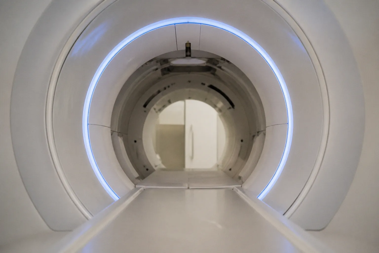
The price difference begins with the detector stack. PET requires scintillation detectors and photon sensors positioned around the patient. MRI requires a high-field magnet, gradient system, RF transmit and receive chains, cryogenic infrastructure, and a room designed around a strong static magnetic field. In a simultaneous system, the PET detector assembly must operate inside that magnetic environment without degrading MRI performance or becoming a safety hazard.
That constrains engineering choices at every layer:
- PET detector materials must tolerate the magnetic field and the electromagnetic environment generated by MRI gradients.
- Detector electronics must avoid injecting noise into the RF receive chain.
- MRI components must not distort PET photon detection or create unacceptable attenuation.
- The gantry must preserve patient access, thermal management, serviceability, and field homogeneity.
- Attenuation correction must account for RF coils, patient positioning, table structures, and other hardware visible to the PET system but not represented cleanly in conventional MR images.
A PET/CT scanner has its own complexity. It also has a more established architecture and a lower capital threshold. CT supplies attenuation information directly through x-ray measurements. MRI does not. PET-MRI therefore replaces a relatively direct attenuation map with a model-derived estimate built from MR data, segmentation, hardware templates, and correction algorithms.
That does not make PET-MRI inferior. It changes the failure modes. A CT attenuation map is exposed to beam-hardening and metal artifacts. An MR-derived map is exposed to tissue classification errors, truncation, motion, coil geometry, and signal voids. The reconstruction pipeline must tolerate missing or ambiguous information. It does not receive a free physical measurement of electron density.
The cost is not limited to the scanner invoice. A simultaneous PET-MRI service requires MRI-compatible monitoring, controlled access, specialized technologists, PET radiopharmaceutical handling, MRI safety screening, and a workflow that does not treat the two modalities as independent appointments. The system must be used intensively enough to justify its fixed cost. Otherwise, its simultaneous capability becomes an expensive idle state.
Simultaneous PET-MRI is not priced as two scanners placed in one room. It is priced as one tightly coupled system whose failure modes also become coupled.
The economic comparison is blunt:
| Parameter | Simultaneous PET-MRI | Sequential PET plus MRI or PET/CT workflow |
|---|---|---|
| Typical capital cost | Approximately $5–7 million | Approximately $1.5–2 million for a 128-detector PET/CT system, with MRI acquired separately when required |
| Spatial relationship between datasets | Acquired in the same examination and coordinate frame | Registered retrospectively across acquisitions |
| Soft-tissue contrast | MRI-derived and highly configurable | MRI provides it only if a separate MRI examination is performed |
| Attenuation correction | Model-based MR attenuation correction | CT attenuation correction for PET/CT; MRI data may remain separate |
| Motion consistency | Can model or observe motion during concurrent acquisition | Patient position and anatomy may differ between examinations |
| Workflow burden | One hybrid protocol, high technical complexity | More scheduling and registration overhead, but more flexible equipment utilization |
| Best technical argument | Temporal and spatial correspondence | Lower cost, broader availability, simpler modality-specific operation |
The table hides one operational truth. Sequential imaging is not a single protocol. It can mean PET/CT followed by MRI, PET followed by MRI on separate days, or PET and MRI performed consecutively in the same institution. These arrangements have different registration errors and different logistics. The comparison must therefore be made against the actual local workflow, not an idealized alternative.
Quantifying spatial precision: why 51% is meaningful, and why it is not enough
The strongest argument for simultaneous PET-MRI is not simply that both images are acquired at the same appointment. It is that anatomy can change between acquisitions. Respiratory phase shifts. The diaphragm moves. Abdominal organs deform. The thorax expands and contracts. Lesions near moving boundaries do not remain in a fixed coordinate system merely because the patient remains on the table.
A study published in Radiology by Brendle and colleagues found that simultaneous acquisition improved alignment of abdominal organs by an average of 51% compared with retrospectively fused PET and MR images. That is a substantial geometric result. It means the hybrid system can reduce a specific source of uncertainty: the mismatch between functional PET signal and anatomical MR structure caused by different acquisition times and respiratory states.
This gain is not equivalent to a 51% improvement in diagnosis. It is not a 51% increase in sensitivity. It is an alignment metric. The distinction is not semantic. Registration accuracy and clinical accuracy are related but non-identical variables.
A better alignment can yield:
- More reliable localization of focal uptake relative to organ boundaries.
- Cleaner interpretation of lesions adjacent to the diaphragm or bowel.
- Reduced ambiguity when PET activity is spatially diffuse.
- More defensible radiotherapy or biopsy targeting in anatomically mobile regions.
- Better temporal correspondence when metabolic activity and morphology are interpreted together.
The benefit becomes more complicated when the patient breathes freely. PET acquisition integrates signal over time. MRI sequences may use breath-holds, respiratory navigators, gating, or different sampling windows. A single “simultaneous” examination still contains multiple temporal scales. The PET data may represent an accumulated distribution. The MR image may represent a narrower respiratory state.
Research on thoracic PET/MRI protocols has reported a 60% alignment improvement when free-breathing or exhaling breath-hold strategies were used instead of inhaling MR acquisition. The specific protocol matters. The scanner does not automatically remove motion. It provides access to synchronized acquisition and motion information. The reconstruction must still use that information correctly.
This is where acquisition physics becomes decisive. The MR sequence may provide a respiratory signal, navigator echo, self-gating signal, or repeated anatomical frame. The PET reconstruction may then be corrected, gated, or registered against an MR-derived motion model. Each step reduces one error while potentially increasing another. Gating decreases temporal blur but reduces effective counts per reconstructed phase. Motion correction improves spatial correspondence but depends on the accuracy of the motion estimate. A poor motion model can create sharp, confidently wrong images.
The attenuation correction problem
MRI signal intensity does not map directly to tissue density. Cortical bone produces little conventional MR signal, while air also appears dark. PET attenuation correction must distinguish them using anatomical priors, ultrashort-echo sequences, atlas information, or other model-based methods. The result is an estimate, not a direct measurement.
The practical consequences are familiar to anyone auditing hybrid images:
1. Bone may be misclassified. This can bias PET activity near the skull, spine, or pelvis.
2. The patient may exceed the MR field of view. Truncated anatomy can compromise the attenuation map.
3. RF coils and table structures may be incompletely represented. Hardware attenuation must be modeled separately.
4. Motion can desynchronize the attenuation map and emission data. The PET reconstruction then combines signals from different anatomical states.
5. Metal and implants remain difficult. MRI artifacts and PET attenuation errors can interact rather than remain isolated.
These are not arguments against simultaneous PET-MRI. They are constraints on what the alignment number means. A precisely aligned PET image with a biased attenuation map is not automatically a quantitatively reliable PET image.
Alignment is a geometric property. Clinical utility appears only when the geometric gain survives attenuation correction, motion modeling, reconstruction, and interpretation.
Diagnostic performance in neuro-oncology: parity is a result
Neuro-oncology is often presented as a natural domain for PET-MRI. MRI supplies high soft-tissue contrast, diffusion, perfusion, spectroscopy, and anatomical detail. PET supplies a molecular or metabolic signal that can complement structural imaging. The head is less affected by respiratory motion than the abdomen. The two datasets can be acquired with the patient in a stable position. The theoretical case is strong.
The clinical evidence is more restrained.
In a study of 38 patients with IDH wild-type glioblastoma, researchers compared simultaneous FET-PET and DSC perfusion MRI with sequential acquisitions. They found no significant difference in diagnostic performance. That finding does not invalidate simultaneous acquisition. It limits the claim. In this cohort and application, the hybrid architecture did not demonstrate a diagnostic advantage over the sequential workflow.
This is the type of result that acquisition teams often mishandle. Technical superiority in synchronization is treated as if it must produce superior clinical classification. It does not. Diagnostic performance is a function of signal contrast, lesion biology, protocol quality, reader behavior, disease prevalence, and the decision threshold. If both workflows already provide adequate spatial correspondence in the brain, the additional simultaneity may have little effect on the final interpretation.
The comparison is especially sensitive to the clinical question:
- Tumor recurrence versus treatment effect: Perfusion, diffusion, amino-acid uptake, and contrast enhancement may all contribute. Better temporal registration can help, but it may not change the classification if the dominant signal is already clear.
- Surgical planning: MRI may remain the primary anatomical dataset. PET can add biological information, but the benefit depends on whether uptake changes the surgical boundary or merely confirms a known abnormality.
- Radiotherapy planning: Registration quality matters, but immobilization, sequence distortion, and contouring practice may dominate the residual uncertainty.
- Longitudinal follow-up: A simultaneous system reduces the chance that the patient’s position and anatomy differ between studies. The benefit is real, but its size depends on how consistently the sequential protocol is executed.
- Research imaging: Simultaneous acquisition may be more valuable because investigators can correlate dynamic signals and reduce timing ambiguity, even when routine clinical diagnosis remains unchanged.
The lack of a significant difference in the glioblastoma study should therefore be read as a boundary condition. If a center’s primary indication is neuro-oncology, the business case cannot rest on the assumption that simultaneous acquisition automatically improves diagnostic performance. The center must identify the specific endpoint. Lesion localization? Quantitative kinetic modeling? Treatment response? Target definition? If the endpoint is not defined, the scanner is being purchased on architecture rather than outcome.
When simultaneity has less leverage
The hybrid advantage weakens when the sequential workflow is already controlled. A patient is positioned consistently. MRI and PET/CT are acquired close together. Registration is performed with deformable algorithms. Respiratory state is documented. The diagnostic question does not depend on sub-organ motion. In that environment, simultaneous PET-MRI may still reduce logistical friction for selected patients, but its image-quality advantage may be marginal.
The advantage also weakens when the MRI portion is not optimized for the clinical indication. A simultaneous system does not make a generic MR protocol equivalent to a dedicated neuroimaging protocol. Gradient performance, diffusion timing, susceptibility behavior, RF coil geometry, and sequence acceleration still determine the data. PET cannot compensate for an underpowered MRI acquisition. MRI cannot repair inadequate PET counts.
Workflow efficiency and the 30-minute threshold
A simultaneous PET-MRI examination is only clinically credible if the acquisition time remains compatible with patient tolerance and department throughput. A practical target is to keep image acquisition within approximately 30 minutes. Longer protocols may be technically richer, but they consume scanner time, increase motion exposure, and reduce the number of patients the system can support.
The 30-minute threshold is not a universal law. It is a workflow constraint. PET counts accumulate over time. MRI sequences compete for the same examination window. Every additional sequence has a cost:
- More diffusion directions increase scan duration.
- Higher spatial resolution reduces signal per voxel or requires stronger acceleration.
- Multiphase contrast imaging adds timing dependencies.
- MR spectroscopy requires shimming, localization, and spectral stability.
- Motion correction sequences consume time without directly contributing to the final diagnostic series.
- Respiratory gating can discard data or extend acquisition.
The protocol designer is therefore balancing several quantities at once: PET count statistics, MRI SNR, temporal resolution, spatial resolution, gradient duty cycle, SAR, patient motion, and reconstruction latency. Improving one variable commonly degrades another.
A high-resolution MRI sequence can increase anatomical confidence while lowering SNR. Parallel imaging can shorten acquisition while amplifying noise and g-factor penalties. Compressed sensing can reduce sampling burden while placing more responsibility on the reconstruction prior. PET reconstruction can apply stronger regularization to produce a cleaner image, but excessive regularization can suppress small or low-contrast lesions.
This is why “workflow efficiency” should not be measured only from patient arrival to patient departure. The relevant metric is usable information per scanner hour. A 25-minute protocol that produces diagnostically adequate PET and MRI data may outperform a 40-minute protocol with more sequences but higher motion burden. Conversely, a short protocol that omits the sequence needed for the actual clinical decision is not efficient. It is incomplete.
The hidden time costs
Simultaneous PET-MRI removes some duplication. The patient does not need to be repositioned between separate scanners. The PET and MRI datasets share a temporal context. But hybrid systems add preparation and recovery steps that ordinary MRI operations may not carry.
The department must manage:
- Radiopharmaceutical administration and uptake timing.
- MRI safety screening and implant verification.
- MR-compatible monitoring for patients who cannot remain unattended.
- Attenuation-map quality control.
- PET detector and MRI subsystem calibration.
- Correction of motion, truncation, and susceptibility artifacts.
- Reconstruction pipelines that may be more computationally demanding than either modality alone.
- Data transfer and archival requirements for large, multi-series examinations.
The scanner can acquire both modalities concurrently. The department still has to interpret, archive, route, and quality-control both data streams.
A sequential pathway can be less elegant and more resilient. PET/CT and MRI can be scheduled independently. A failed MRI sequence does not necessarily invalidate the PET/CT examination. A hybrid protocol may create a single point of operational failure: motion, injection timing, hardware artifact, or attenuation-correction failure can compromise the combined study.
The efficient PET-MRI protocol is not the one with the most sequences. It is the one that preserves the clinical decision while constraining motion, count noise, and scanner occupancy.
Hardware physics: where the premium is actually spent
The simultaneous architecture is a compromise between incompatible physical demands. MRI wants a controlled electromagnetic environment. PET wants sensitive photon detection over a large solid angle. Neither subsystem can be treated as an accessory to the other.
Magnet and gradients
The static magnetic field constrains PET detector technology and electronics. Gradient coils add rapidly changing magnetic fields. Their slew rate and amplitude determine how quickly k-space can be traversed, but they also create electromagnetic interference and acoustic load. The hybrid system must manage these effects without allowing PET electronics to contaminate the MR receive chain.
The MRI side remains governed by familiar trade-offs:
- Higher gradient slew rates can shorten echo spacing and improve diffusion performance, but they increase peripheral nerve stimulation constraints and acoustic burden.
- Stronger gradients can support faster echo-planar readouts, but susceptibility artifacts remain a major limitation near air-tissue interfaces.
- Parallel transmit and receive strategies can improve spatial control, but RF inhomogeneity and SAR remain sequence-dependent.
- Dedicated head or body coils can improve SNR, but they also contribute to PET attenuation and must be represented in correction maps.
The hybrid design does not remove these constraints. It adds another layer of coupling.
PET detector integration
The PET detector assembly must be compact enough to fit within the MRI system while maintaining detection efficiency and timing performance. The detector ring must not create unacceptable attenuation or field distortion. Electronics must operate reliably in the magnetic and RF environment. Thermal management must occur without introducing vibration, noise, or new safety problems.
The outcome is a system whose performance cannot be reduced to a single sensitivity value. PET image quality depends on count density, detector timing, reconstruction, attenuation correction, patient size, uptake distribution, and motion. MRI quality depends on field strength, coil geometry, sequence design, gradient behavior, and reconstruction. Simultaneous acquisition connects these variables through the patient’s motion and the scanner’s correction pipeline.
Reconstruction is part of the scanner
The final image is not a direct photograph of physiology. It is the output of a chain:
- RF excitation and reception.
- Gradient encoding.
- k-space sampling.
- Coil combination.
- Parallel or compressed reconstruction.
- Geometric distortion correction.
- PET coincidence detection.
- Randoms, scatter, and normalization correction.
- MR-based attenuation correction.
- Motion estimation.
- Image registration or motion-compensated reconstruction.
- Quantitative calibration and display.
An improvement at one stage can expose a weakness at another. Better motion estimation can reveal attenuation-map misalignment. More aggressive denoising can reduce visible noise while altering lesion contrast. A sharper reconstruction can make residual registration errors more conspicuous. The image may appear cleaner and become less trustworthy quantitatively.
That is the central acquisition issue. Simultaneous PET-MRI is not valuable because the hardware is integrated. It is valuable when the integration produces a lower total error for the clinical question.
Strategic implementation: when the premium is defensible
The purchase decision should begin with indications that depend on synchronized information, not with the availability of a capital budget.
Simultaneous PET-MRI is easier to justify when several conditions align:
- The service evaluates anatomy that moves or deforms during the examination.
- PET and MRI signals must be interpreted in the same physiological state.
- The patient population benefits from MRI’s soft-tissue contrast or reduced ionizing radiation relative to CT-based imaging.
- The center has sufficient radiopharmaceutical access and MRI/PET technical expertise.
- The protocol can remain near the 30-minute acquisition target.
- The institution can support advanced motion correction and MR attenuation-correction quality control.
- The scanner will serve both clinical and research workloads rather than a narrow indication with low volume.
- Sequential imaging produces recurring registration problems that affect management, not merely image aesthetics.
Abdominal and thoracic applications offer the clearest technical argument because respiratory motion directly degrades retrospective fusion. The reported 51% average improvement in abdominal organ alignment is not trivial. Neither is the reported 60% improvement under specific thoracic acquisition strategies. But these values do not prove superiority across every tumor type, every respiratory protocol, or every patient.
Neuro-oncology requires a more disciplined business case. The head is anatomically stable compared with the thorax. Sequential registration can already be strong. The study of 38 IDH wild-type glioblastoma patients found no significant diagnostic-performance difference between simultaneous FET-PET with DSC perfusion MRI and sequential acquisition. A center buying PET-MRI primarily for glioblastoma follow-up must therefore identify a narrower advantage: reduced appointment burden, improved research timing, quantitative multiparametric modeling, or a specific treatment-planning use case.
What a credible deployment plan contains
A serious implementation plan should specify more than scanner utilization. It should define the failure modes that the system is expected to reduce.
A useful plan contains:
1. An indication-level endpoint. For example, improved lesion localization in respiratory motion, reduced repeat imaging, or better temporal registration in a defined research protocol.
2. A matched comparator. Sequential PET/CT plus MRI, PET plus MRI on the same day, or the actual local standard. The comparator determines the apparent gain.
3. A protocol budget. Time allocated to PET uptake, anatomical MRI, diffusion, perfusion, spectroscopy, attenuation correction, and motion handling.
4. An artifact audit. Truncation, susceptibility, coil attenuation, respiratory mismatch, metal, and failed motion correction should be tracked separately.
5. A quantitative quality-control layer. SNR, uniformity, geometric distortion, SUV stability, and registration error require independent monitoring.
6. A clinical decision map. The team must document where the simultaneous data changed interpretation or management.
7. A utilization model. Capital cost, service contracts, radiopharmaceutical supply, staffing, downtime, and reconstruction infrastructure all affect the premium.
Without these controls, the hybrid scanner risks becoming a prestige installation. It will produce technically sophisticated images. That is not the same as producing better decisions.
Clinical necessity or luxury?
The binary framing is too crude, but the purchasing decision is not. Simultaneous PET-MRI is clinically valuable when temporal and spatial correspondence are themselves part of the diagnostic problem. The 51% abdominal alignment improvement demonstrates that the system can solve a real registration limitation. Thoracic protocols show the same principle under motion-sensitive acquisition conditions.
The evidence does not support a universal claim of diagnostic superiority. In glioblastoma follow-up, simultaneous and sequential acquisition produced similar diagnostic performance in the cited cohort. The additional hardware did not automatically yield a better clinical classification. That result should discipline the discussion, not end it.
The system is a premium instrument for a narrow class of errors: motion, temporal mismatch, soft-tissue localization, and integrated multiparametric research. It is not a replacement for protocol design. It is not a substitute for adequate PET counts, stable MRI SNR, accurate attenuation correction, or competent interpretation.
The capital gap remains severe. At $5–7 million, simultaneous PET-MRI demands a workload and indication set that can sustain the architecture. A $1.5–2 million 128-detector PET/CT system provides a lower-cost, more established route to oncological PET imaging, with MRI added when the clinical question requires it. Sequential imaging is less elegant. It is often more flexible. In controlled workflows, it can be diagnostically equivalent.
The blunt conclusion is this: simultaneous PET-MRI is not a luxury because it is technically extravagant. It becomes a luxury when the institution cannot name the error it will reduce. If respiratory motion, temporal registration, or integrated research acquisition determine the outcome, the premium may be defensible. If the justification is simply that both modalities occupy the same gantry, the physics has not made the case.