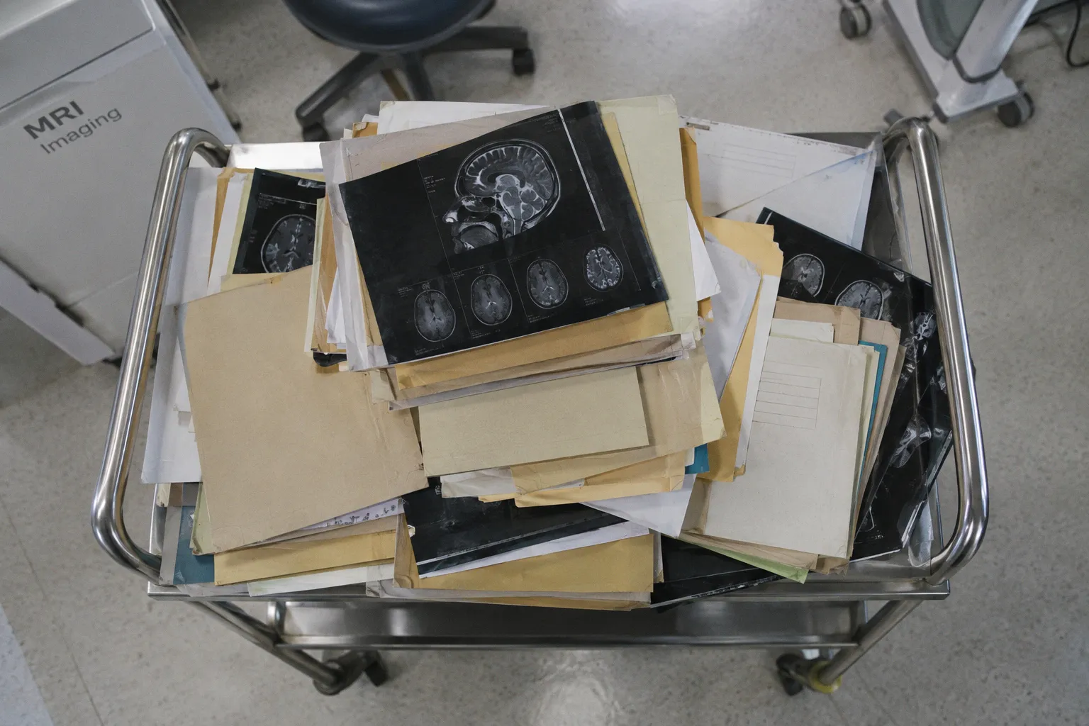
When a multi-site imaging trial encounters a PACS metadata mismatch, the cost often appears first as a line item nobody planned for: 15 to 20 minutes of manual reconciliation per imported study, multiplied across every prior scan, every subject, and every site. That is the visible bottleneck. The less visible cost is the queue it creates around the imaging core lab, site coordinators, data managers, and clinical operations team.
Everything else follows from that queue: delayed image transfers, unresolved queries, repeated requests to sites, missed review windows, and the slow-motion friction between a sponsor and the people responsible for keeping local systems operational.
The DICOM standard is not the villain. It has been stable, versioned, and broadly adopted for decades. The problem is the assumption that “DICOM-compliant” at each site automatically means “DICOM-aligned” with every other site in the trial. It does not. The gap between those two phrases is where multi-center imaging studies lose time, introduce avoidable reconciliation work, and weaken confidence in the dataset.
A PACS metadata mismatch in multi-site clinical trials is rarely one dramatic failure. More often, it is a collection of locally reasonable decisions that become incompatible when the data is centralized.
The root cause: vendor-specific DICOM implementations
When “compliant” stops meaning “compatible”
DICOM is a contract. Each tag—patient ID, study date, accession number, referring physician, institution name—has a defined meaning, a defined value representation, and defined valid inputs. A PACS that follows the contract is, by definition, compliant.
What the contract does not fully specify is how each site should populate those tags for non-standard workflows: clinical trial accessioning, research-only scanners, secondary captures, de-identified exports, or studies imported from outside institutions. At that point, each vendor and each hospital IT team makes a local decision. Local decisions do not automatically aggregate into a clean research dataset.
Consider the (0010,0020) Patient ID field. For routine clinical reads, it may carry the medical record number. For a trial, the same field might need to carry a randomized subject ID, a site-specific screening number, or a de-identified research alias. The standard defines the field’s purpose, but it does not by itself determine which identifier a particular trial must place there.
As a result, Site A may export the MRN, Site B the screening number, and Site C a one-way hash. All three exports can be syntactically valid DICOM. None necessarily resolves to the same subject record in the EDC.
The same divergence appears across the rest of the header:
Study Description (0008,1030)may containBRAIN MR TRIAL VISIT 1at one site andTRIAL01_V1_BRAIN_MRat another.- A third site may leave Study Description blank and rely on the accession number for routing.
Institution Name (0008,0080)may be spelled differently across facilities in the same health system.Referring Physician (0008,0090)may disappear when the order originates from a non-affiliated clinic.- A site may preserve a value in a private tag that another site does not export at all.
- De-identification may remove a field that the trial had assumed would remain available for reconciliation.
None of these choices necessarily violates DICOM. Together, they can break the trial’s data dictionary, matching logic, routing rules, and audit process.
This is the origin point of many downstream mismatches: not a broken standard, but a standard with room for local interpretation at the exact boundary where the trial needs deterministic behavior.
DICOM does not fail at the trial level by itself. The failure usually appears at the boundary between a site’s local interpretation and the trial’s centralized data model.
The metadata problem is semantic, not merely syntactic
A common integration mistake is to validate whether a tag exists without validating what the value means in the trial. A populated Patient ID is not automatically a usable trial identifier. A valid Study Date is not necessarily the date the protocol calls the imaging visit date. A non-empty Series Description does not guarantee that the series can be classified by the central reader or analysis pipeline.
This distinction matters because DICOM headers can pass technical validation while still failing operational validation.
For a research workflow, the relevant questions are more specific:
1. Does the identifier resolve to the correct trial subject without exposing prohibited clinical identifiers?
2. Does the date correspond to acquisition, procedure, export, or upload?
3. Does the value identify a protocol visit, or only describe the clinical order?
4. Can the same logic be applied across vendors, institutions, and repeat scans?
5. Is the value stable after de-identification, import, transfer, and re-export?
A site can answer these questions differently without realizing that it has created a cross-site inconsistency. That is why a generic DICOM conformance statement is not enough for a clinical research integration. Conformance describes capabilities. It does not define the trial’s interpretation of the data.
Technical barriers in Query/Retrieve and DICOM Part 4
What DICOM Query/Retrieve actually returns—and what it does not
Most PACS-to-PACS and PACS-to-research-archive integrations rely on DICOM Query/Retrieve, defined in DICOM Part 4 through the Query/Retrieve Information Model. It is the workhorse of clinical imaging exchange, and for ordinary operational retrieval it works well.
Its limits become more visible when a sponsor needs bulk metadata for a clinical trial imaging audit, site-level reconciliation, or centralized research classification.
Query/Retrieve exposes the attributes supported by the relevant information model and by the PACS implementation. In practice, that often means operational fields such as:
- Patient ID
- Study Instance UID
- Series Instance UID
- Modality
- Study Date
- Accession Number
- Study and Series descriptions
It does not automatically provide every header attribute a research data manager may need. Protocol-specific sequence parameters, reconstruction details, private tags used for local trial flagging, de-identification provenance, and operator-entered comments may be unavailable through the query interface, returned inconsistently, or exposed only through vendor-specific behavior.
If the sponsor needs those values, there are usually two practical choices. The first is to retrieve the full DICOM objects, including pixel data, and parse the headers locally. That provides a broad view of the source metadata but increases transfer, storage, and processing requirements. The second is to negotiate a vendor- or site-specific metadata export, which may be more efficient but can take substantial coordination and may not be portable across sites.
For a daily clinical reading list, Query/Retrieve is generally sufficient. For a sponsor pulling research metadata across many sites, it can become a structural bottleneck.
| Metadata need | DICOM Query/Retrieve | Bulk object parsing | Direct PACS database query |
|---|---|---|---|
| Patient ID, Study UID, Modality, Study Date | Often available through standard query levels | Available in the object header | Available if the site permits access |
| Sequence parameters and reconstruction details | Model- and vendor-dependent | Available when present in the object | Available only if stored and exposed |
| Private tags and local trial flags | Usually vendor-dependent | Available when not removed during export | Site-specific |
| Bulk extraction without retaining pixel data | Not always practical | Possible with header-only processing | Possible, but tightly coupled to the PACS |
| Audit trail of tag modifications | Not inherent in the Q/R model | Available only if provenance is separately logged | Site-specific |
| Portability across institutions | Relatively good at the standard level | Depends on parser and de-identification rules | Usually poor |
The table illustrates an uncomfortable point: the most standard interface is not always the most informative one. A research workflow may need to combine standard retrieval with a controlled metadata-extraction process rather than expecting the PACS query layer to serve as a complete research database.
Why vendor-specific behavior matters
The same DICOM attribute can behave differently depending on where it is generated, transformed, or exported. A scanner may write one value, the modality worklist may supply another, and the PACS may alter or normalize it during ingestion. A de-identification tool may remove or replace fields before the research archive receives the object. A routing rule may match on one attribute at one site and on a different attribute elsewhere.
These transformations are not necessarily errors. They become errors when the trial assumes that the original value, the local value, and the exported value are interchangeable.
A useful integration review therefore follows the object through its actual path:
- scanner or acquisition console;
- modality worklist and RIS;
- local PACS ingestion;
- de-identification or pseudonymization;
- research archive or image-management platform;
- central reader or analysis environment;
- EDC or eCRF mapping layer.
At each handoff, the team should identify which attributes are preserved, which are transformed, which are generated locally, and which are no longer available. Without that map, a site may pass a connectivity test while still failing the trial’s data-integrity requirements.
The hidden cost of manual data reconciliation
The 15-to-20-minute tax per imported study
Here is the math that rarely makes it into the protocol budget.
When a site imports an outside imaging study—a prior scan from a referring hospital, a screening MRI from a satellite clinic, or a follow-up from a patient who has moved—the local PACS may receive DICOM objects whose metadata does not map cleanly to local patient and study identifiers. The MRN in the imported file may not match any local MRN. The accession number may come from a different RIS. The study description may be uninformative. The operator field may be empty. A de-identification step may have removed the field the local routing rule expects.
Someone has to resolve the discrepancy. Usually that person is a radiology technologist, research coordinator, PACS administrator, or imaging data manager. The work can include:
1. Opening the incoming study and confirming its provenance.
2. Identifying the correct local patient or trial subject.
3. Determining whether the study is a new examination, a prior, or a duplicate.
4. Updating demographic and study-level fields.
5. Linking the images to the appropriate local order or research accession.
6. Confirming that the study is routed to the correct archive or review queue.
7. Checking whether the change created a new object, replaced an existing one, or left both versions available.
8. Documenting the reconciliation so that the decision can be explained during monitoring or audit.
In many operational workflows, this reconciliation takes roughly 15 to 20 minutes per imported study. The exact time varies with the quality of the incoming data, the local PACS interface, the number of systems involved, and whether the person handling the case has the authority to correct the record.
The important point is not the precise duration. It is the repeated nature of the work. A single difficult study is manageable. The same ambiguity repeated across prior scans, subjects, and sites becomes a standing operational dependency.
A trial does not budget only for the scan. It also pays, somewhere in the workflow, for every ambiguous identifier that arrives with it.
Why reconciliation is more than an administrative nuisance
Manual correction introduces a second problem: the correction itself becomes part of the data history.
If a coordinator changes a Patient ID, accession number, or study description in the local PACS, the central system may see only the corrected value. If the original value is not retained in a controlled audit trail, it becomes difficult to reconstruct what arrived, who changed it, why the change was made, and whether the same object was already transferred elsewhere.
This matters for both clinical interpretation and research analysis. A duplicate may be mistaken for a repeat visit. A corrected accession number may look like a new acquisition. A missing series may be attributed to the scanner when it was actually filtered during routing. A subject mismatch may remain undetected if the corrected record happens to match an existing patient.
The reconciliation process should therefore be treated as a data-transformation step, not just a clerical task. At minimum, the trial workflow needs a way to record:
- the incoming identifiers;
- the normalized trial identifiers;
- the reason for the change;
- the person or system that made the change;
- the date and time of the change;
- the destination systems affected by the correction;
- whether the images were retransmitted after correction.
Without that information, a trial can end up with apparently clean records and weak provenance.
Strategies for standardizing metadata for EDC and eCRF integration
From the DICOM header to the structured research record
The central problem in multi-center imaging data integration is not moving the files. PACS can push, pull, route, and archive DICOM objects across institutional boundaries with reasonable reliability. The harder problem is mapping the contents of the DICOM header into the structured fields an Electronic Data Capture system expects in the electronic Case Report Form.
The eCRF may require a protocol visit, acquisition date, modality, laterality, sequence category, subject identifier, and imaging status. The DICOM object may contain several dates, multiple identifiers, free-text descriptions, and site-specific values. Those representations do not become equivalent merely because both systems can store text.
An eCRF can also accept a null or an apparently valid value without making the underlying inconsistency obvious. The discrepancy may surface only during a data review, a monitoring visit, a central image read, or database lock—when the cost of fixing it is measured in queries and protocol deviations rather than minutes.
A robust mapping strategy needs at least three layers.
1. A trial-specific identifier strategy
The same subject at Site A and Site B must resolve to the same research record without relying on a local MRN. The trial should define which identifier is used for research exchange, where it is stored, and how it is protected during de-identification.
The identifier strategy should also distinguish among:
- screening identifiers;
- randomized subject identifiers;
- local patient identifiers;
- imaging accession numbers;
- object- or study-level UIDs;
- pseudonyms used by a central archive.
Those values may all be useful, but they are not interchangeable. A mapping table should state which one is authoritative for subject matching and which ones are retained only for provenance or local operations.
2. A controlled naming convention
The protocol should define how Study Description, Series Description, Protocol Name, and accession-related fields are used for trial classification. Free text is not automatically unusable, but it is expensive to normalize after the fact.
A workable convention should account for repeat scans, unscheduled visits, screening failures, withdrawals, and outside studies. It should also specify what happens when the local PACS cannot enforce the preferred value. If the fallback is not defined in advance, every site will invent one.
3. A data structure that matches the EDC
The DICOM attributes selected for EDC mapping must correspond to the eCRF data dictionary in meaning, format, and null handling. The review should cover:
- field length;
- value representation;
- permitted character set;
- date and time semantics;
- expected units;
- repeatability;
- missing-value codes;
- whether a field is required, optional, or conditionally required;
- whether the value is copied from DICOM or derived by a transformation rule.
A field can fail even when its text looks correct. A date may be stored in a format the ingestion layer does not parse. A private tag may contain a value that is meaningful only at one site. A sequence parameter may be present but expressed in units that the eCRF does not expect. These are radiology workflow data integrity issues, not cosmetic formatting problems.
Standardize before export, not during rescue
The most reliable place to enforce a trial convention is before the images leave the site. If the research archive has to infer the correct subject, visit, or protocol from ambiguous metadata, the central team is already operating in recovery mode.
That does not mean every PACS must be rebuilt. It may be enough to establish a controlled export profile, a research routing rule, or a site-side transformation service that creates a trial-ready copy while preserving the clinical source. The key is to define the transformation once and apply it consistently.
A useful division of responsibilities looks like this:
| Workflow layer | Primary responsibility | Typical control |
|---|---|---|
| Acquisition and scheduling | Capture the correct protocol and visit context | Worklist values and scanner protocol naming |
| Local PACS/RIS | Preserve or apply site-approved identifiers | Routing rules and export profiles |
| De-identification | Remove or transform protected identifiers predictably | Versioned profiles with test cases |
| Research archive | Validate required metadata before acceptance | Ingestion rules and quarantine queues |
| EDC/eCRF | Store structured trial variables | Data dictionary and controlled mappings |
| Imaging core lab | Resolve exceptions and monitor patterns | Reconciliation log and trend review |
The purpose is not to force every institution to use identical internal workflows. It is to ensure that the output crossing the trial boundary has predictable semantics.
Ensuring data provenance through configuration alignment
The three network values that gate the pipeline
Before strategic metadata mapping can work, the basic DICOM network configuration must be correct. Communication between a PACS, research archive, imaging core lab, or ingestion node depends on the receiving endpoint being correctly identified and reachable.
Three values commonly appear in the initial configuration:
| Configuration value | What it controls | Common failure mode |
|---|---|---|
| Application Entity (AE) Title | The DICOM application identifier used by the endpoint | A mismatch in the routing table leads to association rejection or an endpoint that does not recognize the caller |
| Port number | The TCP port on which the endpoint listens for DICOM traffic | A firewall, service, or port mismatch produces connection refusal or a timeout |
| IP address or hostname | The network location used to reach the endpoint | VPN, NAT, DNS, or routing problems prevent the request from reaching the intended system |
These values are often described as if they were a simple three-item checklist. In practice, they sit inside a larger chain that includes firewall rules, VPN routes, DNS behavior, TLS requirements where applicable, calling and called AE validation, storage commitment settings, and the receiving system’s acceptance policy.
A configuration error can produce a clear technical error. The DICOM association may be rejected. The connection may be refused. A request may time out. A C-STORE operation may return a status that the sending system records in its logs. These are not inherently silent failures.
The risk is different: the error may be visible at the DICOM or network layer but overlooked at the trial-workflow level. A site may see a failed transfer in a local log without creating a corresponding operational exception. The sponsor may see no image in the central repository but have no immediate indication whether the cause was a rejected association, a routing rule, a missing study, or a delayed export.
That is how a logged technical failure can become an apparently silent data-loss event for the trial.
A network error can be perfectly visible to the PACS and still be effectively invisible to the trial unless the workflow turns it into a tracked exception.
Test the complete path, not just the connection
A successful C-ECHO or basic connectivity test proves only that two DICOM endpoints can communicate at that moment. It does not prove that the correct study will be selected, exported, accepted, indexed, de-identified, and mapped to the right research subject.
Pre-go-live testing should use representative test objects and should exercise the full path:
1. Send a test study from the intended source system.
2. Confirm that the receiving endpoint accepts the association.
3. Confirm that the images and metadata arrive.
4. Check whether the expected series are present.
5. Verify the subject and visit mapping.
6. Confirm that de-identification has behaved as intended.
7. Check the archive’s indexing and search behavior.
8. Confirm whether the EDC or eCRF receives the expected structured values.
9. Review the sender and receiver logs.
10. Record the test result in a site-level configuration record.
The test set should include more than a clean, ideal examination. It should include an outside prior, a repeat visit, a missing optional field, a de-identified object, and any modality or sequence type that the trial expects to analyze. This is where DICOM tag inconsistency in research usually becomes visible—before real subjects depend on the workflow.
Configuration drift is a provenance problem
Even a correctly configured site can drift out of alignment. PACS upgrades, network changes, vendor migrations, new de-identification profiles, scanner replacements, and changes to local routing rules can alter the behavior of an established integration.
For that reason, configuration management belongs inside the trial’s imaging governance rather than being treated as a one-time IT task. The sponsor or imaging core lab should maintain a current record of:
- sending and receiving AE Titles;
- approved ports and network endpoints;
- system owners and escalation contacts;
- de-identification profile versions;
- expected metadata mappings;
- test-study results;
- change dates and approvals;
- known site-specific exceptions.
The aim is not bureaucratic completeness. It is to preserve the chain of provenance from acquisition to analysis. If a study arrives with an unexpected value, the team should be able to determine whether that value came from the scanner, the local PACS, a transformation service, the de-identification step, or a later manual correction.
What actually fixes a multi-site imaging workflow
The practical fixes are not glamorous, but they are concrete.
Stop treating compliance as compatibility. Each site needs a trial-specific DICOM conformance and metadata review before the first subject is enrolled. The review should cover actual sample objects, not only a generic statement that the PACS supports DICOM.
Define the authoritative identifiers. Decide which value identifies the trial subject, which value identifies the imaging visit, and which values are retained for local operations or provenance. Do not leave those decisions to individual site coordinators.
Standardize the export boundary. Where possible, enforce the trial’s naming and mapping rules before data leaves the site. If a site requires a transformation service or a research-specific export profile, document it and test it.
Use Query/Retrieve for what it does well. It remains useful for operational discovery and retrieval. Do not assume it is a complete bulk research metadata interface. For audit and reconciliation work, plan for header parsing, controlled exports, or another agreed method of obtaining the required attributes.
Turn technical failures into workflow exceptions. Association rejection, connection refusal, timeouts, and storage errors should create actionable records linked to a site, study, subject, and attempted transfer. A log entry that nobody reviews is not a functioning control.
Retest after meaningful change. A PACS upgrade or de-identification-profile change can alter the trial data path even when the site believes the integration is unchanged.
Preserve the correction history. Manual reconciliation may be unavoidable, but it should be traceable. The original value, corrected value, reason, and downstream impact belong in the provenance record.
None of this appears in a vendor brochure. It is distributed across imaging operations, site IT, clinical data management, the imaging core lab, and trial oversight. That distribution is precisely why it is easy to assume that someone else owns the problem.
The standard is not the whole solution, and a successful connection is not proof of a successful workflow. A multi-site trial produces reliable imaging data when the local PACS implementations, network configuration, metadata rules, de-identification behavior, and EDC mappings are treated as one chain rather than as separate technical tasks.
The fix is unglamorous and entirely achievable: define the meaning of each identifier, test the real transfer path, monitor the errors that systems already record, and preserve enough provenance to explain every correction. That work costs less before the first scan than after the dataset has reached central review—or database lock.