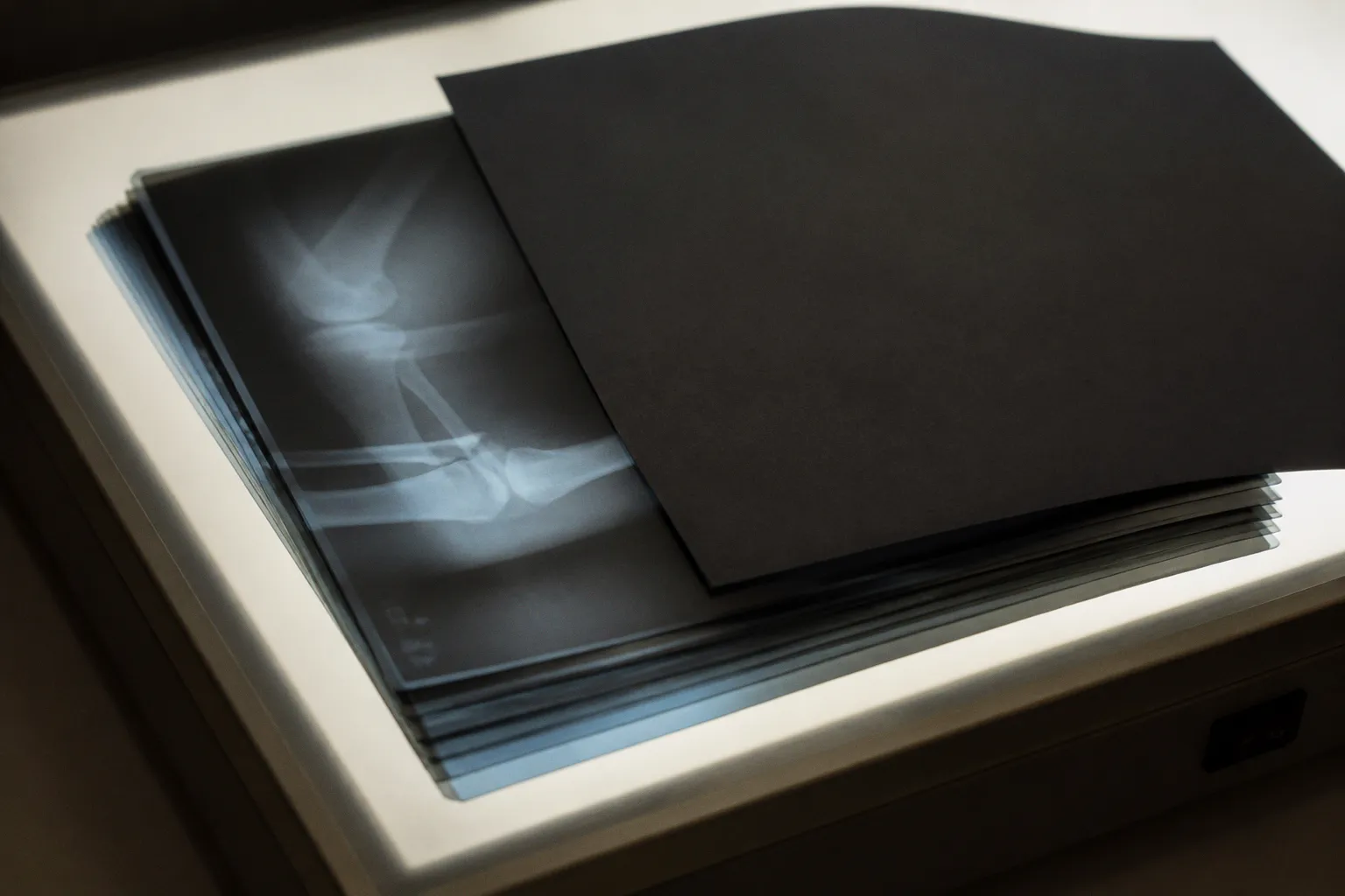
In structural brain MRI, the risk does not end when a patient name is removed from the DICOM header: the scan may preserve enough facial anatomy to support three-dimensional reconstruction, while private tags, accession numbers, dates, or device-specific fields can continue to link the study record to an individual.
This is the central difficulty of MRI data de-identification for research. It is not a single export command and not a setting that can be enabled once at the edge of a PACS. It is a workflow in which metadata sanitization, pixel-level processing, validation, access control, and longitudinal traceability have to agree with one another. If one layer is treated as sufficient, the clinical trial may appear compliant while remaining vulnerable in precisely the places that routine review is least likely to examine.
Beyond the Header: The Dual-Layer Anonymization Mandate
DICOM carries more than the visible clinical description of an examination. A study may include patient identifiers, dates, accession information, referring physician details, institution names, device information, study descriptions, operator fields, and private vendor tags. Some of these elements are immediately recognizable as protected health information. Others become identifying only when combined with the trial database, a radiology information system, a scheduling record, or the structure of a longitudinal imaging series.
For this reason, DICOM anonymization standards are best understood as a set of controlled transformations rather than a universal definition of “clean.” The metadata layer answers one question: what identifying fields remain in the DICOM objects and their associated files? The pixel layer answers another: can the patient be recognized from the image content even after the header has been sanitized?
A three-dimensional brain MRI makes the second question unusually important. Structural sequences may retain facial contours, orbital regions, nasal structures, and other anatomical features that are sufficient for facial reconstruction. Skull-stripping or defacing can reduce that exposure, but these operations introduce their own scientific and operational considerations. A defaced image may be appropriate for a particular research analysis while being unsuitable for another, especially when the study depends on anatomy near the skull, scalp, orbit, or cranial boundary.
The workflow therefore begins by defining what the dataset must preserve. A trial focused on cortical thickness may tolerate a different pixel-level transformation from a study examining craniofacial anatomy, surgical planning, or subtle extracerebral signal. The research question is not an administrative detail; it determines whether anonymization can be aggressive, whether an original image must remain in a protected source system, and how investigators will document the transformation.
Removing the patient name from a DICOM header is a necessary step, but for structural brain MRI it is not the definition of de-identification.
A useful clinical trial workflow separates the source image from the research derivative. The source examination remains governed by the clinical imaging environment, where the patient identity and care context are required for treatment, reporting, audit, and lawful re-identification. The research copy is created through a controlled process, assigned a study-specific identifier, and moved into the research environment only after metadata and pixel-level checks have been completed.
That separation also protects longitudinal research. A trial may need to connect several MRI examinations from the same participant without exposing the participant’s name or medical record number. The solution is not to retain the original patient identifier in the research repository. It is to generate a controlled pseudonymous key, maintain the linkage in a restricted location, and ensure that the research identifier is consistent across approved time points without becoming a public identifier in its own right.
Consider the implications for an enterprise imaging platform. If a DICOM object is de-identified before it leaves the clinical domain, the process must preserve enough information for the research viewer, analysis pipeline, and audit trail to function. If de-identification occurs after the file has already been copied into a broad-access cloud bucket, the technical transformation may be correct while the overall exposure has already occurred. Timing, location, and permissions are part of the de-identification design.
Navigating DICOM PS 3.15 Annex E and Metadata Sanitization
DICOM PS 3.15 Annex E provides a structured basis for deciding how metadata elements should be handled through Application Level Confidentiality Profiles. Its action codes describe the treatment applied to individual attributes, including:
- D — replace: substitute a value with a dummy or otherwise non-identifying value;
- X — remove: delete the attribute;
- K — keep: retain the value because it is necessary and considered acceptable within the profile;
- C — clean: preserve the element while removing identifying content within it;
- X/D — remove or replace: apply the appropriate action depending on the attribute and the implementation context.
These codes are useful because they move the conversation away from vague assurances that a file has been “anonymized.” They create a map between the DICOM attribute, its clinical or technical purpose, and the transformation expected during export. The map should be versioned, reviewed, and tested against the actual objects produced by the scanners and routing systems in the trial.
In practice, this is where many projects become more complicated than their original protocol suggests. DICOM conformance does not mean that every implementation populates every field in the same way. A vendor may use private tags for acquisition details; a scanner console may write a study description that contains a protocol nickname; an intermediary gateway may add routing information; a PACS export may preserve fields that were absent in the original transmission. The research team needs to validate the final dataset, not merely approve the configuration that was intended to create it.
The Patient Name tag, (0010,0010), and Patient ID tag, (0010,0020), are obvious examples, but they are only the beginning. Dates can become identifying when combined with a rare diagnosis, a recruitment record, or a known visit schedule. Accession numbers and study identifiers can provide a bridge back to clinical systems. Institution and operator fields may reveal the site or staff member involved. Free-text fields are particularly difficult because their content may not follow a predictable pattern and may contain identifiers entered by a human operator.
A robust metadata review should therefore ask several connected questions:
- Which attributes are required for image reconstruction, quality control, or sequence harmonization?
- Which values are needed to preserve the timing of longitudinal visits, and can they be transformed without retaining the original date?
- Are private tags being removed, retained under a documented exception, or copied into a controlled research field?
- Could an accession number, study UID, or series UID be used to connect the research file to a clinical system?
- Are derived objects, segmentation files, reports, and screenshots being processed under the same policy as the original images?
- Does the output remain internally consistent after identifiers are replaced or regenerated?
The last question is easy to underestimate. A research pipeline may require stable relationships between a study, its series, and individual instances. De-identification can replace identifiers while preserving those relationships, but the replacement process must be deliberate. A file that has no obvious patient name but contains broken or duplicated UIDs may create downstream errors, encourage manual workarounds, or cause researchers to retrieve the wrong object from the source system.
The checklist should describe decisions, not only tags
A useful clinical trial DICOM workflow records the reason for each retained element. “Keep” should not mean “the tool left it untouched.” It should mean that the project has established why the element is necessary, who can access it, and how the risk is managed.
The record should include at least:
1. The input boundary. Identify where the image enters the research workflow: scanner, PACS, vendor gateway, clinical research platform, or a separate export workstation.
2. The object scope. Include original DICOM images, enhanced multi-frame objects, secondary captures, derived images, segmentation objects, dose or protocol records, and any accompanying reports that may contain identifiers.
3. The transformation profile. Document the DICOM PS 3.15 Annex E profile or equivalent project-specific policy, including the action applied to private tags and free-text fields.
4. The identifier strategy. Define how patient, study, series, and instance identifiers are replaced, and where any re-identification key is held.
5. The validation method. Specify which automated tests and human reviews confirm that the output matches the approved profile.
6. The exception process. Record what happens when a scanner, site, or software version produces an unexpected attribute or a value that cannot be removed without damaging the research use case.
This kind of documentation is not bureaucratic padding around the real science. It is what allows a multi-site study to distinguish a controlled exception from an unrecognized leakage path.
Addressing the 18 HIPAA Safe Harbor Identifiers
HIPAA Safe Harbor defines the removal of 18 categories of identifiers, including patient names, geographic subdivisions smaller than a state, most elements of dates other than the year, telephone and fax numbers, email addresses, medical record numbers, health plan identifiers, account numbers, certificate or license numbers, vehicle and device identifiers, web URLs, IP addresses, biometric identifiers, full-face photographs and comparable images, and other unique identifying numbers or codes.
For MRI research, two features deserve particular attention. First, dates are not simply another group of header fields. Exact examination dates can be essential for modeling disease trajectory, treatment response, or interval change, yet they can also expose a participant when combined with recruitment or clinical records. A trial may need to transform dates into relative study time, shift them consistently for a participant, or retain only the year, depending on the protocol and governance decision. The correct choice depends on the scientific design and the applicable privacy framework; it should not be left to the default behavior of a general-purpose DICOM utility.
Second, the Safe Harbor category for full-face images has a direct relationship to structural MRI. A volumetric brain scan is not a conventional portrait, but facial anatomy can still be represented in the pixels. The fact that the image is stored as medical data does not eliminate the reconstruction risk. In other words, satisfying a header-oriented de-identification routine does not automatically resolve the image-content question.
This is where a “HIPAA-compliant MRI de-identification” claim needs careful wording. HIPAA compliance is not established by a software brand, a green status indicator, or a single successful export. It depends on the covered entity’s or research organization’s policies, implementation, risk analysis, access controls, contractual arrangements, and validation. A tool can support a compliant workflow; it cannot, by itself, make every workflow compliant.
The same restraint applies when a study operates across jurisdictions. HIPAA Safe Harbor is a specific U.S. method, while other regimes may use different definitions, safeguards, or expectations around pseudonymization, biometric information, and research reuse. A global trial should not assume that a Safe Harbor-oriented DICOM profile is a universal privacy standard. The metadata policy, pixel policy, consent language, and governance process need to be reviewed together.
A privacy rule can tell the workflow what to remove; it cannot decide what the research question can afford to lose.
Mitigating Facial Reconstruction Risks in Structural MRI Datasets
Metadata stripping and facial anonymization solve different problems. The first changes the information attached to the image. The second changes the information contained in the image. Treating them as interchangeable is one of the most consequential mistakes in MRI data de-identification for research.
Pixel-level anonymization commonly includes defacing or skull-stripping. Defacing removes or alters facial regions that may support recognition, while skull-stripping attempts to retain intracranial structures and remove non-brain tissue. Neither operation should be treated as universally harmless. A mask can remove relevant anatomy, create edge artifacts, alter intensity distributions near the boundary, or complicate registration and quality control. The choice must be evaluated against the sequences and analyses included in the protocol.
A practical assessment begins with the dataset’s anatomy and not with the name of the software. T1-weighted structural scans are an obvious focus, but other 3D acquisitions, reformatted series, derived volumes, and exported screenshots may also preserve recognizable facial structure. If the dataset contains multiple versions of the same examination, anonymizing one series while leaving an unprocessed derivative available can defeat the intent of the process.
The study team should define:
- Which modalities and orientations require defacing or skull-stripping;
- Whether the transformation is applied to the original research export or to a derivative;
- How image quality is assessed after processing;
- Whether facial removal affects the endpoints, registration, segmentation, or morphometric measurements;
- How failures are handled when the algorithm does not produce a satisfactory mask;
- Whether the processed output and the unprocessed source are stored in separate access zones.
Visual review remains useful, particularly during pipeline validation and when onboarding a new scanner or sequence. Automated checks can confirm that the expected mask exists, that image dimensions and orientation remain consistent, and that processing completed without an error. They cannot always determine whether a facial contour has been sufficiently removed or whether the transformation has damaged a clinically relevant region.
The validation set should include difficult cases rather than only clean examples. Motion, incomplete coverage, unusual head positioning, implants, susceptibility artifacts, variable field of view, and sequences acquired with different protocols can all affect pixel-level processing. A multi-site trial should also test across scanner manufacturers and software versions, because the same nominal sequence name does not guarantee identical image geometry or metadata behavior.
There is a scientific tension here that deserves to remain visible. The more anatomy a study removes, the smaller the residual identification risk may become, but the greater the possibility that the research signal near the removed region will be altered. The answer is not to choose privacy or scientific validity as an absolute. It is to define the minimum image content required for the approved analysis, preserve the original clinical source under restricted controls, and make the transformation reproducible enough that its consequences can be studied.
Evaluating De-identification Toolkits: Why Default Settings Fail
The ecosystem of DICOM and neuroimaging utilities includes tools such as Clinical Trials Processor, DICOMCleaner, PyDICOM with Deid, and the RSNA Image Share De-Identifier. These tools can be valuable components in a clinical trial pipeline, but their presence in a workflow does not remove the need for configuration and verification.
A study evaluating 10 non-commercial DICOM toolkits found that only one successfully de-identified all 50 target PHI elements under default settings, while only five achieved full de-identification after manual configuration. The finding is important not because it identifies a universally best tool, but because it demonstrates the gap between software availability and workflow assurance. Default settings are often a starting point, not evidence that the output satisfies the project’s policy.
Several failure modes recur across implementations:
Private tags remain outside the assumed policy
Private tags are not automatically harmless because they are not part of the most visible standard fields. They may contain device, institution, acquisition, or workflow information, and their content can differ by vendor and software version. The project must decide whether to remove them wholesale, retain a documented subset, or transform them under a validated rule.
Free text is harder to govern than fixed fields
A structured field can be mapped to a defined action. A free-text field may contain a name, a location, a clinical comment, or an internal code entered in an unexpected format. A header review that checks only a list of common tags may miss identifiers embedded in descriptions or notes.
Secondary and derived objects are overlooked
The original series may be clean while a secondary capture, reformatted image, segmentation object, or analysis export retains the original study information. The research inventory should include every object that can leave the clinical environment, not only the files most commonly opened by the imaging viewer.
Pixel data is treated as an administrative field
A DICOM toolkit may correctly process metadata while making no claim about facial reconstruction. This is not a defect in the tool; it is a boundary of the problem. The trial’s policy must explicitly connect metadata de-identification with defacing or skull-stripping when structural MRI is involved.
Re-identification links are stored too broadly
A pseudonymous study ID can support longitudinal analysis, but the linkage table that connects it to the clinical identity is itself sensitive. It should not travel with the research dataset, sit in the same unrestricted object store, or be accessible to every analyst who needs the images.
A more dependable evaluation process uses representative exports from every participating site and scanner configuration. The team should inspect raw DICOM headers, process the files using the candidate profile, and then compare the result against a target inventory. The same review should be repeated after routing through the actual PACS, cloud gateway, or research imaging platform, because intermediary systems can add or transform attributes.
| Layer | What the workflow must examine | Typical evidence of completion |
|---|---|---|
| Standard metadata | Patient name, patient ID, dates, accession and study identifiers, institution and operator fields | Automated tag report compared with the approved profile |
| Private metadata | Vendor-specific fields and implementation-defined attributes | Private-tag inventory, documented removals or exceptions |
| Relationships between objects | Study, series, instance, frame, and derived-object identifiers | Consistent regenerated identifiers without unintended source linkage |
| Pixel content | Facial anatomy in 3D structural MRI and other relevant image types | Defacing or skull-stripping review, including difficult cases |
| Longitudinal linkage | Stable research identity across visits without exposing clinical identity | Restricted linkage service or separately controlled key |
| Output environment | PACS, research repository, cloud viewer, analysis workspace, backups | Access logs, permissions review, and confirmation that only approved derivatives are exposed |
The final evidence should be reproducible. A reviewer ought to be able to determine which input was processed, with which profile and software version, under what date and operator context, and what validation result was produced. This is especially important when a trial runs for years and the imaging estate changes underneath it.
Integrating De-identification with Enterprise Imaging Workflows
De-identification is often designed as though the file exists in isolation. Clinical imaging does not work that way. MRI data moves through scanners, modalities worklists, PACS, vendor-neutral archives, research gateways, cloud storage, viewers, analysis platforms, and sometimes external sites supporting tele-radiology or centralized reading. Each transition can preserve, add, transform, or expose identifying information.
A reliable architecture assigns a clear responsibility to each boundary. The clinical PACS remains the authoritative source for patient-linked imaging. A research export service creates the derivative according to the approved DICOM profile. The research repository receives only the transformed objects, while the clinical-to-research linkage is held separately. The cloud-based MRI viewer should authenticate against the research identity rather than silently restoring the clinical patient context through an integration link.
This does not mean that interoperability must be abandoned. DICOM remains essential for imaging objects, while HL7 and FHIR-based services may support orders, scheduling, consent, trial enrollment, or controlled exchange of clinical context. The privacy question is whether those interfaces carry more information than the research use requires. A research viewer may need a study identifier, acquisition time relative to baseline, modality, and series description. It may not need the patient’s legal name, full birth date, clinical account number, or direct link to the hospital chart.
The same principle applies to automated pipelines. If an analysis service receives files through an API, the service should be designed around the pseudonymous research identity and the minimum necessary metadata. Logs, temporary files, thumbnails, error reports, and backups deserve review because they can preserve identifiers after the primary DICOM object has been cleaned.
A clinical trial can document this as a chain of custody:
1. The clinical system produces the source examination and records its origin.
2. A controlled service applies metadata and pixel-level transformations.
3. Automated validation checks the resulting DICOM objects.
4. Human review examines representative images and exceptions.
5. The research repository receives only approved derivatives.
6. Analysis tools use research identifiers and retain auditable processing records.
7. Re-identification, when legitimately required, occurs through a restricted clinical or study-governance process rather than an analyst-facing database.
The chain is most valuable when it includes failure handling. What happens if a new scanner software version adds an unrecognized private tag? What happens if a defacing algorithm fails on a particular sequence? Does the system block export, quarantine the object, or allow it through with a warning? A warning that no one is required to resolve is not a control.
A Verification Culture for Longitudinal MRI Research
The hardest part of de-identification is rarely the first successful export. It is preserving the same standard across time, sites, software updates, protocol amendments, and secondary analyses. Longitudinal studies amplify small inconsistencies because an identifier leak or processing change can affect every visit associated with a participant.
The validation process should therefore be treated as a living part of the imaging protocol. When a new acquisition sequence is introduced, its metadata profile and pixel-level behavior need review. When the PACS or cloud platform changes, the team should test whether object relationships, private fields, and audit information are preserved as intended. When a new analysis requires anatomy that was previously removed, the study must revisit the balance between scientific need and residual identification risk rather than quietly distributing an unprocessed copy.
This shift allows us to see de-identification as part of data quality, not an obstacle placed in front of data quality. A dataset with clean identifiers but inconsistent visit linkage is not operationally safe. A dataset with excellent longitudinal structure but unreviewed facial anatomy is not privacy-complete. A dataset that passes a header script but cannot be traced to a defined transformation profile is difficult to defend when questions arise.
The most useful checklist is consequently concise in principle, even when the implementation is detailed:
- The project has defined whether it needs de-identification, pseudonymization, or both.
- The approved DICOM profile identifies what is removed, replaced, cleaned, or retained.
- Private tags and free-text fields have been included in testing.
- The policy covers derived objects and secondary captures as well as original images.
- Structural MRI pixel data has been assessed for facial reconstruction risk.
- Defacing or skull-stripping has been selected in relation to the scientific endpoint.
- Research identifiers support longitudinal analysis without exposing the clinical identity.
- The re-identification key is segregated from the research image repository.
- Validation uses representative data from each site, scanner, and relevant software path.
- Export is blocked or quarantined when an unexpected field or processing failure appears.
- The process is documented well enough to reproduce the decision months or years later.
No checklist can eliminate judgment. It can, however, make the judgment visible.
For clinical MRI research, the safest approach is neither maximal removal nor faith in a default toolkit. It is a layered method that follows the image from its clinical origin through every research boundary, asks what information the biology truly requires, and verifies that the resulting object is safe in both its header and its pixels. The deeper purpose is not simply to satisfy a privacy rule. It is to preserve the participant’s dignity while allowing the research record to remain coherent enough to reveal change over time—the subtle degradation, treatment response, or preserved cognitive reserve that longitudinal neuroimaging is uniquely positioned to describe.