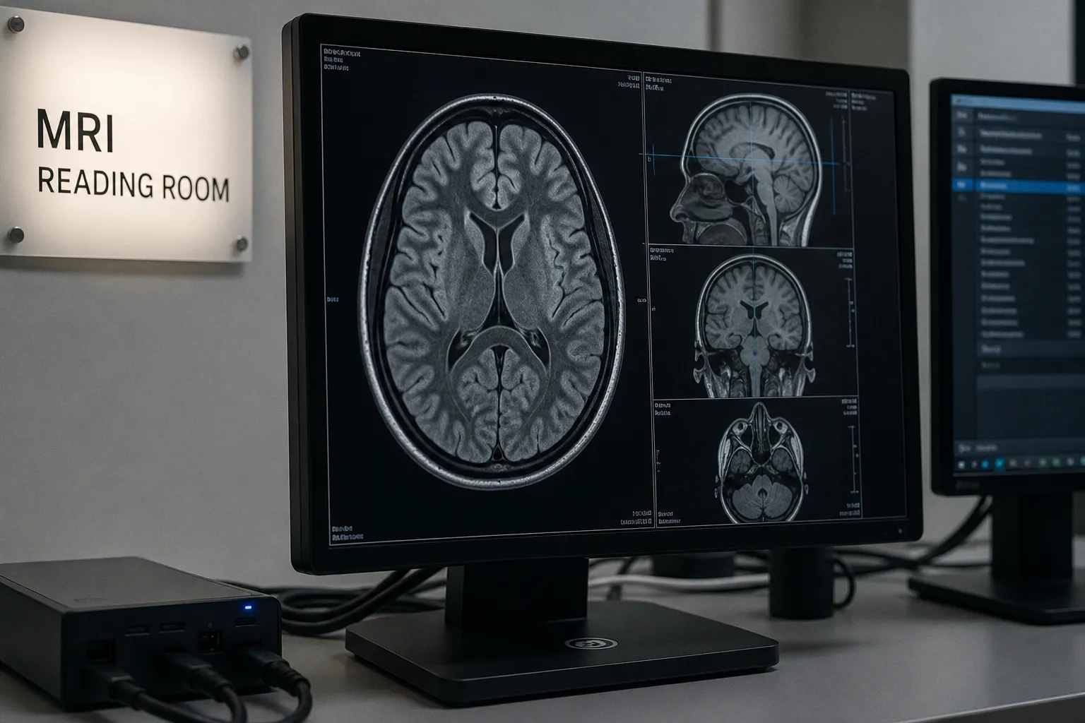
The difference is rarely explained by the word cloud alone. It is determined by where DICOM parsing, decompression, window/level mapping, and image rendering actually occur: on the server, inside the browser, or across a carefully balanced combination of both.
This is the central engineering dilemma behind cloud MRI viewer rendering latency. A viewer designed for a stable workstation with a capable GPU may behave very differently on a thin clinical laptop, a teleradiology connection, or a hospital network carrying several concurrent studies. Consider the implications for enterprise imaging: rendering architecture is not merely a question of interface smoothness. It shapes how quickly a radiologist can reach the relevant series, how reliably a longitudinal study can be reviewed, and how much infrastructure must remain available when imaging volumes continue to grow.
MRI data growth is already substantial, with datasets increasing at an annual rate of about 21% in the cited MICCAI research data, while functional MRI grows even faster. That trajectory changes the meaning of a practical workflow. A viewer that performs well with a handful of compact studies may become an operational constraint when it must serve high-resolution examinations, prior comparisons, multiplanar reconstructions, and remote users at the same time.
The rendering decision begins with the DICOM object
A DICOM study is not simply a stack of ordinary image files. It carries pixel data together with metadata describing acquisition parameters, orientation, spacing, patient and study identifiers, modality information, and the relationship between individual instances and series. MRI adds further complexity through varying contrasts, diffusion-weighted acquisitions, susceptibility-sensitive sequences, perfusion data, and examinations assembled from multiple series with different spatial and temporal characteristics.
A cloud MRI viewer therefore has to solve several problems before the image becomes clinically useful:
- retrieve the correct instances from PACS or an enterprise imaging archive;
- preserve the geometry and metadata required for anatomical navigation;
- decode compression formats that may not be natively supported by the browser;
- apply window and level transformations without losing the interpretive value of the source data;
- stream enough of the study to support rapid review while additional images continue loading;
- maintain a secure relationship between the viewer, identity provider, archive, and clinical information systems.
The location of these operations defines the architecture.
With server-side rendering, the backend parses DICOM, decompresses or transforms the pixel data, applies rendering logic, and sends browser-friendly frames to the client. These may be delivered as JPEG or PNG images through a REST API or over a WebSocket connection. The browser is primarily responsible for presentation, interaction, and display rather than for understanding every native detail of the DICOM object.
With client-side rendering, the browser receives more of the original imaging data and performs a greater share of decoding and visualization locally. WebGL can use the client GPU for rapid pixel operations, while WebAssembly allows high-performance C or C++ compression libraries to run within the browser, including codecs needed for formats such as JPEG 2000 and JPEG-LS.
Neither approach is universally superior. The relevant question is where the clinical workload should be placed, and whether the network, browser, and backend can sustain that decision under real operating conditions.
Cloud MRI viewer performance is governed less by where the image is stored than by where the expensive work is performed.
DICOMweb and the shift from study download to image streaming
The older mental model of medical image viewing assumes that a workstation retrieves a complete study and then opens it locally. That model is increasingly uncomfortable in a cloud environment. A full MRI examination can contain many series, and the radiologist often needs only a small portion of the study immediately: perhaps the axial T2-weighted sequence, a diffusion series, or a prior examination for side-by-side comparison.
DICOMweb changes the conversation by treating retrieval as a web-native operation. WADO-RS, the RESTful service for obtaining DICOM instances and related resources, supports progressive access to imaging data. Instead of waiting for an entire study to arrive, the viewer can request images or frames as they become relevant to the reading task. In the cited performance description, progressive WADO-RS streaming enables diagnostic-quality images to appear in under two seconds rather than requiring more than ten seconds for a complete study download.
That difference is clinically meaningful, even when the absolute numbers vary between environments. The first image establishes orientation. The first few slices help the radiologist decide whether the selected series is the one needed. The ability to begin interpretation while the remainder of the study continues to arrive is particularly valuable in emergency workflows and in remote reporting, where a long pause can interrupt the cognitive sequence of image review.
DICOM introduced server-based rendering capabilities to WADO-RS in 2015. The significance of that addition is architectural: a web client does not have to receive every original pixel and perform every transformation itself. It can request a rendered presentation from the server, reducing the burden on the browser and making the retrieval path more predictable for devices with limited local compute.
This does not remove the need for thoughtful DICOM integration. The viewer still has to communicate with the archive, understand study and series relationships, and preserve the metadata required for navigation and audit. In an enterprise setting, it may also sit between PACS, a radiology information system, identity services, and clinical applications using HL7 or FHIR-based interfaces. The image may be rendered correctly and still fail as a workflow tool if the radiologist cannot reach it from the relevant worklist or if patient context is not carried safely across the session.
Server-side rendering: control at the backend
Server-side rendering for MRI places the most demanding parts of the imaging pipeline in a controlled environment. DICOM parsing, codec support, pixel conversion, and often window/level processing occur on backend infrastructure before frames are transmitted to the browser.
The practical advantage is consistency. The organization can define the compute environment, monitor its resource use, patch the relevant libraries centrally, and avoid relying on the capabilities of every endpoint. A browser that cannot natively decode JPEG 2000 does not need to do so if the backend has already converted the data into a browser-friendly representation. This is one reason server-side rendering remains attractive for zero-footprint DICOM viewer architecture, especially when the viewer must operate across a mixed fleet of clinical devices.
A server-side pipeline can also be easier to align with centralized governance. De-identification may occur before data reaches a research workspace. Access controls can be enforced at the service boundary. Audit events can be associated with requests for studies and rendered frames. In regulated clinical environments, those controls are not decorative additions around the viewer; they are part of the system’s ability to explain who accessed which data and through what authenticated workflow.
The trade-off is that the backend now carries the burden of rendering sessions. Every active user may require image retrieval, decompression, transformation, and delivery. GPU-enabled infrastructure can improve capacity for some workloads, but it also adds operational complexity and cost. The system must be designed for peaks rather than only for the quiet average: morning worklists, emergency examinations, multidisciplinary review, and simultaneous teleradiology sessions can create very different demand patterns.
Network conditions still matter. Server-side rendering reduces the amount of computation required from the client, but it does not make distance disappear. Frames must travel from the rendering service to the browser, and interaction can feel delayed if each change in slice position or window/level requires a round trip. A server-side architecture therefore benefits from caching, efficient frame requests, sensible image prioritization, and a rendering protocol that does not turn every small interaction into a heavyweight transaction.
The best server-side systems do not simply render everything on demand. They distinguish between what must be available immediately and what can be prepared progressively. The first diagnostic frame may deserve priority. Additional slices can follow. Secondary series and priors can be prefetched according to the reading pattern without forcing the browser to download the full examination before the first useful image appears.
Where server-side rendering is strongest
Server-side rendering is particularly compelling when:
- endpoints vary widely in GPU capability, memory, and browser support;
- the organization needs centralized control over codecs and rendering libraries;
- sensitive datasets should remain within a managed processing boundary;
- the viewer must support older or constrained clinical devices;
- the enterprise wants a uniform behavior across hospitals, reading rooms, and remote reporting locations.
It becomes less comfortable when interaction requires constant, high-frequency manipulation of large image volumes and the network path cannot sustain the resulting exchange. That limitation is not a verdict against the model. It simply means that server-side rendering should be evaluated as an interactive system, not only as a mechanism for producing static images.
Client-side rendering: moving interpretation closer to the radiologist
Client-side rendering transfers more of the imaging work into the browser. This can create a very responsive experience because operations such as window/level adjustment, scrolling, and certain visual transformations do not need to wait for a server response each time. Once the required pixel data is available locally, the interaction can continue even when the connection is no longer carrying every display operation.
The browser’s GPU is central to this model. WebGL fragment shaders can perform the conversion from 16-bit source values to the 8-bit display representation used for visualization. In the cited benchmark, WebGL-based window/level mapping reached 60 frames per second, while CPU-based Canvas 2D rendering struggled at around 12 frames per second for the same DICOM images. The comparison illustrates an important point: the browser is not inherently a weak imaging environment. With the right computational path, it can handle demanding transformations at a speed that feels appropriate for clinical navigation.
But the capability is conditional. A client-side DICOM viewer depends on the endpoint’s GPU, browser implementation, available memory, codec support, and network connection. It also requires a reliable way to decode formats that browsers do not understand natively. WebAssembly addresses part of this problem by allowing optimized compression libraries to execute directly in the browser, including support for non-native formats such as JPEG 2000 and JPEG-LS.
This architecture can reduce repeated server work. The backend may deliver pixel data once, after which the browser performs several local transformations. For a user who is rapidly changing window levels or moving through a volume, that local responsiveness can be valuable. The server is no longer required to generate a new image for every small adjustment.
The price is a larger client responsibility. The browser must receive and hold more data, manage memory carefully, and protect the session against leakage or unintended persistence. A viewer that performs elegantly on a modern workstation may become unstable on a low-memory laptop when several high-resolution series are open simultaneously. The same browser tab that feels instantaneous with one study may begin to stutter when prior examinations, fused modalities, and multiple viewports compete for GPU and memory resources.
There is also a clinical distinction between the ability to render quickly and the ability to render faithfully. A high frame rate is useful only if the transformation preserves the intended intensity relationships, spatial information, and metadata-driven presentation. Speed cannot compensate for an imprecise implementation of window/level behavior or for a workflow that makes it difficult to determine which series, orientation, and acquisition parameters are currently visible.
Server-side and client-side rendering compared
| Parameter | Server-side rendering | Client-side rendering |
|---|---|---|
| DICOM parsing | Performed in backend infrastructure | Performed partly or substantially in the browser |
| Codec handling | Centralized on the server, with browser-friendly output | Requires browser support or WebAssembly codecs |
| Window/level interaction | May require new server-rendered frames | Can be performed locally on the GPU |
| Endpoint requirements | Lower and more predictable | Higher, especially for GPU, memory, and browser capability |
| Network dependence during interaction | Greater when transformations require round trips | Lower after sufficient pixel data is loaded |
| Infrastructure burden | Concentrated in rendering services | Distributed across user devices |
| Governance and control | Easier to centralize | Requires careful client security and lifecycle management |
| Scaling challenge | Concurrent rendering sessions and backend capacity | Bandwidth, data delivery, and endpoint variability |
| Best fit | Mixed devices and centrally managed workflows | Capable workstations and highly interactive visualization |
The table should not be interpreted as a choice between centralized reliability and local performance in absolute terms. Modern viewers increasingly use hybrid strategies: the server handles retrieval, authorization, and difficult codec operations, while the client uses WebGL for display transformations that benefit from immediate interaction.
A 60 FPS viewer is not automatically a better clinical viewer; the useful measure is how faithfully and predictably it supports the radiologist’s next decision.
Progressive loading is a workflow design, not only a transport feature
Radiology viewer streaming protocols are often discussed in terms of bandwidth, but the more important question is what the user can do while the study is loading. A progressive viewer establishes a sequence of clinical usefulness.
The first visible image should not be selected merely because it is the smallest object. It should help the radiologist orient within the examination. Subsequent frames should support the next likely action: scrolling through the current series, opening another plane, comparing with a prior, or reviewing a specialized sequence. This requires coordination between DICOMweb retrieval, viewer state, and the underlying archive.
WADO-RS can support this pattern by allowing the client to request individual instances or frames rather than treating the study as a single indivisible download. The viewer can prioritize the active series, request additional frames concurrently, and defer less urgent content. When the initial diagnostic image appears in under two seconds, the system has not merely improved a benchmark. It has shortened the interval between opening the case and forming an anatomical understanding of the problem.
Concurrency is another part of the equation. In performance testing of a cloud DICOM API, increasing concurrent image retrieval requests from six to twenty improved frame rates from 30 FPS to 60 FPS for 0.5 MB uncompressed DICOM images within the same region. This result should be read narrowly. It demonstrates the potential effect of request concurrency under the tested conditions; it does not establish a universal performance law for every archive, network, or deployment. The outcome will vary with geographic distance, service limits, cache behavior, image size, compression, and the ability of the backend to process requests in parallel.
Over-aggressive concurrency can also create its own failure mode. A viewer that requests too many images at once may compete with other clinical traffic, exhaust browser resources, or cause the archive to spend effort on images that the radiologist never opens. The architecture must therefore distinguish between parallelism that improves the active reading path and parallelism that simply increases background load.
A mature workflow usually benefits from several layers of prioritization:
1. Open the active study quickly. Retrieve the series and frames needed to establish orientation without waiting for every associated object.
2. Continue the current reading path. Once the first image is displayed, stream adjacent slices or frames in the order the user is likely to inspect them.
3. Prepare likely comparisons. Prior examinations and related series can be requested when the current interaction suggests they will be needed, rather than treating all content as equally urgent.
4. Defer low-priority data. Secondary objects, unused reconstructions, and rarely opened series should not compete with the diagnostic viewport.
5. Respect the infrastructure boundary. Concurrency should be tuned against archive capacity, regional network conditions, and the number of simultaneous readers.
This shift allows performance engineering to become more closely aligned with clinical cognition. The goal is not to move the largest possible volume of bytes. It is to make the next meaningful image available at the moment it supports interpretation.
Integration determines whether the viewer belongs in clinical practice
A cloud MRI viewer can render beautifully and still fail to improve radiology workflow optimization if it exists as an isolated destination. Clinical users do not experience PACS, DICOMweb, RIS, identity management, and reporting tools as separate engineering domains. They experience a sequence: a study appears on a worklist, the patient context is opened, the relevant images are reviewed, priors are compared, and the interpretation is documented in the appropriate system.
That sequence places demands on integration beyond image retrieval.
PACS and enterprise imaging platforms must expose studies in a way the viewer can navigate without ambiguity. Series descriptions, accession information, acquisition dates, laterality, and orientation all contribute to the reader’s confidence that the correct data is being examined. If the viewer’s display labels are incomplete or inconsistent with the archive, a technically fast interface may still impose cognitive friction.
The radiology information system provides another layer of context. A viewer launched from a worklist should preserve the intended patient and study scope while preventing accidental drift into an unrelated examination. Identity and authorization services must support appropriate access without turning every series change into a disruptive authentication event. In larger environments, this may involve single sign-on, role-based permissions, session expiration, and auditable access records.
Research and clinical workflows also diverge in their treatment of identity. A clinical viewer may need identifiable information to support care, while a research workspace may require de-identification before data is made available for analysis. That separation should be reflected in the data path and not left to the user’s memory. De-identification, access controls, and auditability are workflow properties as much as security properties because they determine whether the same platform can safely serve both longitudinal patient care and translational research.
HL7 and FHIR integrations can help connect imaging events and clinical context, but interoperability is not achieved by naming a standard. The implementation must preserve the relationships that matter in practice: which patient, which encounter, which order, which accession, and which study. A viewer that retrieves the correct pixels but loses those relationships creates uncertainty precisely where clinical work requires continuity.
Choosing an architecture for latency, scale, and clinical reality
The most useful architecture is usually not selected by asking whether the organization prefers server-side or client-side rendering in the abstract. It is selected by mapping the reading environment to the computational and network constraints.
A high-volume enterprise with diverse endpoints may favor a server-heavy design because centralized decoding and rendering provide more predictable behavior. A specialist neuroradiology service using modern workstations may place more responsibility on the client to support fluid manipulation of large datasets. A distributed health system may use a hybrid model, keeping authorization, retrieval, and difficult codecs under backend control while allowing WebGL to handle local display operations.
The decision can be framed around several practical tensions:
- Latency versus bandwidth: server-side rendering can reduce the amount of data sent to the browser, while client-side rendering may require more initial transfer but reduce repeated interaction traffic.
- Centralized control versus endpoint capability: backend processing is easier to standardize, while client rendering can make better use of powerful GPUs already present in reading rooms.
- Predictable governance versus local responsiveness: a managed server pipeline simplifies central policy enforcement, while local rendering can make window/level and navigation feel immediate.
- Infrastructure cost versus operational resilience: server rendering concentrates costs in shared services; client rendering distributes the workload but increases variability across devices.
- Performance versus data minimization: sending only rendered frames may limit exposure at the client, while richer client-side workflows may require more pixel data to be present in the browser.
These are not purely technical trade-offs. They influence whether a radiologist can compare a subtle enhancement pattern across time, whether a neurologist can review diffusion findings without interruption, and whether a researcher can follow a patient trajectory across longitudinal imaging sessions. The smallest delay may not matter in isolation, but repeated friction changes how a study is read and how confidently findings are connected to prior examinations.
This is why benchmark numbers need clinical interpretation. A 60 FPS result for WebGL fragment shaders is valuable evidence that GPU-based window/level mapping can support smooth interaction. A frame-rate improvement from 30 to 60 FPS after increasing concurrent requests shows that transport strategy can matter. An initial load under two seconds demonstrates the value of progressive streaming. None of these figures, on their own, guarantees a good viewer. They describe components of a system whose final behavior depends on image size, codec, network path, browser state, archive performance, and the sequence of actions taken by the reader.
The practical case for hybrid rendering
In many cloud MRI deployments, hybrid rendering is the most clinically sensible direction because it recognizes that different operations have different requirements.
The server is well positioned to manage DICOMweb access, authentication, authorization, metadata, de-identification boundaries, and codecs that cannot be reliably handled by every browser. It can also provide server-based rendered presentations when the endpoint is constrained or when a predictable output is preferable.
The client, meanwhile, is well positioned to perform interactions that benefit from immediate feedback. WebGL can apply window/level transformations efficiently on the local GPU, and WebAssembly can extend browser support for specialized compression formats. Once the relevant data is available, the user can often manipulate the image without asking the backend to redraw every state.
A hybrid architecture still requires discipline. It should be clear which representation is authoritative, when pixel data may be cached, how browser memory is released, and which operations must be recorded for audit. It should define graceful degradation: if WebGL is unavailable, does the viewer fall back to a server-rendered stream; if the network weakens, can the reader continue with already retrieved images; if a codec fails locally, can the backend transcode the required series?
These fallback paths are not edge-case polish. Clinical software is used across environments that are rarely uniform for long. Browsers update, laptops are replaced, networks are rerouted, and a remote reading location may have very different conditions from the main hospital. A viewer that remains useful when one layer is limited is more valuable than one that is exceptionally fast only under ideal conditions.
The deeper principle is that rendering should follow the clinical task. Rapid orientation may favor server-rendered progressive frames. Intensive manipulation of already loaded data may favor client-side GPU processing. Large-scale integration and governance may require a centralized service boundary. The architecture earns its place by supporting the complete trajectory from study retrieval to interpretation, not by winning a single synthetic benchmark.
Cloud MRI viewer rendering latency is therefore best understood as a systems problem with a human endpoint. DICOMweb and WADO-RS determine how images move. Server-side rendering determines how much work can be centralized. WebGL and WebAssembly determine what the browser can do with local data. Concurrent requests and progressive loading determine whether the first useful image appears before attention begins to drift. PACS, RIS, identity, and security integrations determine whether the viewer can be trusted inside the workflow at all.
The right design does not promise that every study will open instantly or that one rendering model will suit every institution. It creates a dependable relationship between image contrast, computational location, network behavior, and clinical action. This shift allows software engineering to serve something more enduring than speed alone: the radiologist’s ability to recognize a subtle change, place it on a longitudinal trajectory, and make a decision while the relevant biological signal is still clear.