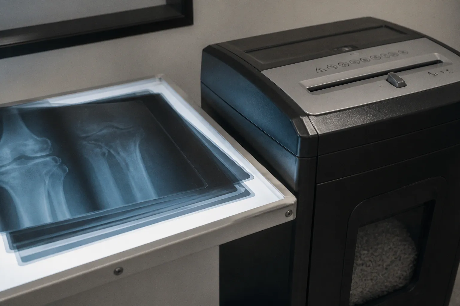
The image may show only a brain MRI, yet the accompanying header can contain a patient name, accession number, study dates, institution, referring physician, device details, unique identifiers, and other fields that make the examination traceable.
That is why a reliable DICOM metadata anonymization workflow for clinical research is not simply a matter of deleting the patient-name tag. It is a controlled transformation of the dataset: removing direct identifiers, managing indirect identifiers, preserving relationships across longitudinal scans, and checking that information has not been embedded in the pixels themselves. The central difficulty is not achieving an empty header. It is creating a dataset that is genuinely safer to share without destroying the biological and temporal structure that makes the research possible.
The anatomy of a DICOM header—and why one visible name is only the beginning
A DICOM file contains two related but distinct forms of information. The first is the viewable pixel data: the actual MRI slices, reconstructed images, diffusion maps, or derived parametric volumes. The second is a structured metadata header made up of hundreds of DICOM attributes, commonly called tags.
Some tags identify the patient directly. Others identify the examination, the institution, the scanner, the study episode, or the technical conditions under which the image was acquired. Many are not obvious to someone opening the series in a clinical viewer. A research export may therefore appear clean at the level of the displayed image while remaining identifiable through combinations of header fields.
Consider the difference between these two risks:
- A patient name is a direct identifier that can usually be removed or replaced.
- A rare examination date, institution name, accession number, and persistent study identifier may together preserve a link back to the source record even after the name has disappeared.
- A series description may contain free text entered by a technologist or radiologist, and free text is difficult to govern with a simple fixed list of tags.
- A device serial number or station name may expose the clinical site or make scans from the same subject easier to connect.
- A burned-in annotation inside the pixel data is not affected by header scrubbing at all.
This layered structure is why automated de-identification of MRI headers must be treated as a workflow rather than as a single software command. Automation is valuable, particularly when a research repository contains thousands of images, but automation only performs safely when the project has already decided what must be removed, what may be transformed, and what needs to remain available for analysis.
In neuroimaging, that distinction becomes especially important because the research value of a dataset often depends on relationships that are not visible in one image. Longitudinal dates may be needed to understand disease trajectory. Subject age and sex may be relevant to biological interpretation, and in some PET-related workflows, demographic information can contribute to calculations such as standardized uptake values. A dataset stripped indiscriminately of all contextual information may be private in a narrow sense but scientifically unusable.
The goal of de-identification is not to erase the study’s history; it is to remove the patient’s identity while preserving the history the science genuinely needs.
Start with the intended research use, not the anonymization button
The correct transformation depends on what the dataset is meant to support. A teaching file, a cross-sectional machine-learning dataset, a longitudinal neurodegeneration study, and a multi-site clinical trial do not have identical privacy or data-utility requirements.
Before exporting a series from PACS, the research team should describe the intended downstream use in operational terms. Will investigators compare scans from the same participant over time? Will images be linked to clinical outcomes? Will multiple institutions contribute data? Will the dataset be used for model training, segmentation annotation, radiomics, or visual review? Each answer changes the metadata that should be retained and the relationships that must survive anonymization.
A practical planning discussion usually covers several questions:
- Which fields are required to identify a participant inside the approved research environment, and which of those fields should be replaced by a study-specific code?
- Does the project need exact acquisition dates, relative intervals, or only the order of visits?
- Must the same subject, study, series, and instance relationships remain intact?
- Are scanner model, field strength, sequence parameters, or acquisition site necessary for the analysis?
- Will the dataset be shared outside the originating institution, and if so, under what access controls?
- Are there local institutional tags or private creator elements that the standard profile may not fully describe?
- Does the image type create a risk of text being burned into the pixel data?
This is where regulatory frameworks and technical standards meet, but they should not be conflated. HIPAA and GDPR establish privacy and data-governance obligations in their relevant contexts. DICOM PS 3.15 Annex E, originating from Supplement 142, provides standardized Attribute Confidentiality Profiles and defined actions for handling DICOM attributes. A profile can guide the technical transformation, but it does not replace the project’s data-governance decision or institutional review process.
The phrase “compliant anonymization” is therefore incomplete unless the workflow defines what compliance means for the specific dataset, jurisdiction, study protocol, and sharing arrangement.
Applying DICOM PS 3.15 Annex E without flattening the dataset
DICOM PS 3.15 Annex E is useful because it moves de-identification away from informal tag lists and toward standardized confidentiality profiles. Rather than treating every attribute as either safe or unsafe, the profiles describe actions that can be applied to fields according to their privacy sensitivity and their role in the imaging workflow.
Supplement 142 and related de-identification rules reference more than 200 header attributes. In protocols associated with the Oxford Medical Imaging Database, for example, 222 tags are referenced in the context of strict de-identification rules. The exact number relevant to a given project may vary with modality, implementation, private elements, and the chosen profile, but the larger point is consistent: the privacy surface is much broader than the handful of fields that a viewer displays by default.
The operations are not all equivalent. Depending on the attribute and the profile, a field may be:
| Operation | What it does in practice | Why it may matter to research |
|---|---|---|
| Remove or null | Deletes the value or leaves the field empty | Appropriate when the attribute has no scientific or technical role |
| Dummy replacement | Inserts a non-zero-length substitute value | Preserves a required structural field without retaining the original identity |
| Encrypt | Transforms the value so it can be recovered under controlled conditions | Useful when a trusted custodian must retain a reversible link |
dateShift | Moves dates according to a defined transformation | Preserves relative chronology while reducing exposure of real calendar dates |
cryptoHash | Derives a consistent transformed value from the original | Can preserve matching relationships without exposing the original identifier |
| Substitute | Replaces the original with a controlled value | Supports a project-specific pseudonym or coded representation |
| Perturb | Alters a value within a defined strategy | May reduce precision while retaining useful distributional information |
refreshUID | Generates new unique identifiers | Breaks links to the original DICOM environment while preserving internal relationships |
The action code itself is less important than the rule governing its use. A date shift that is random for every file can destroy the order of visits. A shift applied independently to each series can make one examination appear to occur on different days depending on the export unit. A UID refresh that generates unrelated identifiers for images that belong to the same series can break reconstruction and annotation workflows.
This shift allows us to see anonymization as a transformation of relationships, not merely a deletion of values. When an MRI study contains repeated scans, the anonymized data must still communicate which images belong to the same participant and which series belong to the same examination—unless the protocol intentionally removes those relationships.
The distinction between anonymization and coded research data
Research teams often use the word anonymization for several different arrangements. A truly irreversible dataset may have no permitted route back to the original participant. A coded or pseudonymized dataset may replace direct identifiers with a study code while retaining a controlled re-identification key elsewhere. Those are not interchangeable models.
A PACS export may therefore require two separate identifiers:
1. An internal research subject identifier that remains consistent across approved longitudinal visits.
2. New DICOM UIDs that no longer expose the original imaging system’s identifiers but remain internally coherent within the exported dataset.
The first supports study design. The second supports technical integrity and separation from the source archive. Treating both as the same problem often produces avoidable errors. A subject code should not be casually copied into every DICOM UID field, and a refreshed UID should not be mistaken for a research participant identifier.
A dependable workflow from PACS export to research repository
The most defensible process has distinct stages, with a clear boundary between the clinical archive and the research environment. The exact software architecture may differ—on-premises service, secured workstation, cloud pipeline, or enterprise imaging platform—but the logic should remain visible and auditable.
1. Define the data contract
Start with a written description of the fields and relationships the study needs. This is not a generic list of “useful metadata.” It should specify the actual variables required for the analysis.
For a longitudinal MRI study, the contract might preserve:
- A study-specific subject code.
- Relative visit order or shifted acquisition dates.
- Modality, body region, and sequence identity.
- Acquisition parameters needed for harmonization or quality control.
- Scanner and site information if the analysis models acquisition effects.
- Relationships among study, series, image instances, and derived objects.
- Demographic attributes required by the approved protocol.
Everything else should have a documented reason to remain. That reason can be technical, scientific, or operational, but it should not be an assumption inherited from the clinical archive.
2. Export into a controlled staging area
DICOM objects should pass through a controlled staging environment before they reach the shared research repository. The staging area is where the original export can be quarantined, transformed, validated, and either released or rejected.
This separation matters because it limits the number of systems that handle identifiable material. It also makes it possible to compare the pre- and post-transformation states without placing the original files into the research workspace.
The workflow should preserve an audit record of what was processed, which profile was used, when the transformation occurred, and whether any files failed validation. The audit record should not itself become a second store of patient identifiers. Operational logging needs the same discipline as image storage.
3. Apply a profile and a project-specific policy
Use the relevant DICOM confidentiality profile as the baseline, then add a documented project policy for local private tags, free-text fields, derived objects, and any attributes that the study must retain.
Private elements deserve particular attention. They may contain vendor-specific acquisition information, reconstruction details, or identifiers that are not covered by a simple public-tag rule. Removing every private element can damage reproducibility; retaining all private elements can preserve information the team did not intend to share. The decision should be made by reviewing the actual scanner and PACS implementations represented in the dataset.
The same applies to free text. A field labelled as a description can contain protocol information in one department and a patient reference in another. A robust workflow therefore combines known-tag rules with a review of how local systems populate those fields.
4. Preserve longitudinal structure deliberately
Time is one of the most important forms of information in clinical neuroscience. The interval between baseline and follow-up may be more useful than the calendar date itself, particularly when studying subtle degradation, treatment response, or cognitive reserve across a disease trajectory.
A dateShift operation can preserve relative timing while reducing exposure of the original dates, but only if the shift is applied consistently according to the study design. The transformation may need to be stable for all dates belonging to one participant, or stable across a defined study, depending on the protocol. If the shift changes from one image to the next, the resulting dataset may contain impossible chronology.
This is also where the difference between “remove all dates” and “protect dates” becomes clinically meaningful. Eliminating dates may appear safer, but it can erase the longitudinal structure required to interpret progression. The right question is not whether dates are present; it is whether their retained form is proportionate to the scientific purpose and governance model.
5. Refresh UIDs while maintaining internal consistency
DICOM UIDs link studies, series, instances, and related objects. An anonymization process should normally create new identifiers that are not tied to the source archive, while ensuring that all references within the exported dataset continue to resolve.
A refreshed UID strategy should be tested against:
- Multi-series examinations.
- Derived images and source-image references.
- Segmentation objects and measurement data.
- Registration and transformation objects.
- Annotation workflows.
- Duplicate exports of the same study, if the research system expects stable matching.
- Re-import into the target PACS or cloud-based MRI viewer.
The last point is easy to overlook. A dataset can be private and internally valid yet fail when loaded into the destination platform because relationships were not retained in the form that the viewer expects.
6. Validate the result at more than one level
Validation should not end when the anonymizer reports success. A useful review has at least three layers.
Metadata validation examines the transformed header for direct identifiers, institutional identifiers, private elements, unexpected free text, and values that should have been removed or altered. It should also confirm that required research fields remain available.
Structural validation checks whether the DICOM objects remain readable and whether the study-series-instance hierarchy is intact. It should include references between images, segmentations, measurements, and derived volumes where those objects are part of the project.
Visual validation opens representative images and looks for identifiers burned into the pixels. Header anonymization does not protect against text that is part of the image itself. This problem is especially familiar in modalities such as ultrasound, but no export pipeline should assume that MRI images are automatically free of embedded labels. Pixel masking or redaction may be necessary when annotations are present.
A small sample is not enough if the repository combines different scanners, locations, operators, or acquisition protocols. The validation set should represent the variation in the source environment, including unusual series and older studies.
The balance between privacy and scientific utility
Overzealous de-identification is often described as the safer choice, but in a research setting it can create a different kind of failure: the data remain technically present while the scientific signal has been severed.
Removing all dates can prevent longitudinal analysis. Removing age and sex can complicate biological interpretation. Stripping acquisition parameters may make it difficult to understand scanner-related variation or harmonize protocols. Deleting all private elements can remove reconstruction information needed to reproduce a processing result. Breaking subject-level identifiers can prevent investigators from distinguishing repeated scans from independent observations.
None of this means that research utility should override privacy. It means that privacy rules should be applied with enough biological understanding to distinguish identity-bearing information from clinically meaningful context.
The most useful way to make that distinction is to classify fields according to their function:
- Direct identity fields identify a person or a clinical episode and generally require removal, replacement, or controlled transformation.
- Linkage fields connect images across studies, visits, or systems and may need stable substitution rather than deletion.
- Temporal fields describe when acquisition occurred and may need date shifting instead of removal.
- Technical fields describe the scanner, sequence, reconstruction, or acquisition environment and may be essential for quality control or harmonization.
- Institutional fields identify a hospital, department, workstation, or operator and should be reviewed for both privacy and site-level disclosure.
- Free-text fields require special handling because their content is not reliably predicted by the tag name.
- Derived-object references preserve the relationship between source images and analyses and should not be changed without testing the complete object graph.
This classification turns a broad privacy exercise into a series of decisions that can be reviewed by clinical, technical, and governance stakeholders.
A dataset is not made research-ready by having fewer tags; it is made research-ready when every retained tag has a reason to exist and every removed tag has been handled without breaking the study.
Why a single anonymization tool is rarely the whole workflow
Clinical imaging environments are assembled from systems that were not always designed as one continuous research pipeline. PACS, radiology information systems, vendor consoles, research databases, annotation platforms, and cloud storage may each interpret DICOM metadata slightly differently.
An anonymizer may correctly process public tags but leave site-specific private elements untouched. A PACS may export a derived object differently from an original acquisition. A cloud viewer may display a field that another system ignores. An annotation platform may rely on identifiers that the research policy treats as disposable. These are integration problems, not merely software defects.
The workflow should therefore be tested across the systems that will actually handle the data:
1. The clinical source produces the export.
2. The staging service transforms the DICOM objects.
3. The validator inspects metadata, structure, and pixels.
4. The research repository receives the approved dataset.
5. The viewer or annotation platform opens and interprets it.
6. Downstream analysis software reads the fields required for processing.
If a project uses HL7 or FHIR-based clinical integration alongside DICOM imaging, the linkage between systems deserves separate review. A clean DICOM header does not automatically make an associated clinical record safe to share. Conversely, a research identifier used in a clinical interface should not expose the original patient identifier simply because both systems need to refer to the same examination.
This shift allows us to treat interoperability as part of privacy engineering. Every bridge between PACS, RIS, research databases, and cloud services is also a possible route by which identity, dates, or institutional information can travel.
Performance, scale, and operational failure modes
Large imaging repositories introduce a practical constraint: de-identification must run consistently enough to support real research operations. Processing benchmarks from integrated workflows have been recorded at approximately 10 images per minute, with some larger jobs requiring around two hours. These figures should be treated as implementation-dependent rather than as a universal performance promise. File size, compression, network design, pixel redaction, validation depth, and the number of objects in each study can all change throughput.
At scale, the most serious operational risk is often not slow processing but silent inconsistency. A pipeline may handle routine brain MRI studies correctly while failing on:
- Multi-frame objects.
- Secondary captures.
- Derived maps.
- Segmentation and measurement objects.
- Studies containing unusual private tags.
- Images with embedded annotations.
- Duplicate or repeated exports.
- Corrupted or incomplete instances.
- Series whose descriptions contain unexpected free text.
Every failed object needs a disposition. It should be quarantined for review rather than passed through because most of the study succeeded. The exception report should explain whether the problem involved an unsupported object type, a missing required attribute, a malformed header, an unresolved reference, or a suspected identifier.
A research team should also define what happens when a participant withdraws consent or when the source clinical record is corrected. If the research dataset is irreversibly anonymized, removal may not be technically possible through the original patient identifier. If the project uses a controlled coded model, a trusted key may allow the data custodian to locate and remove the relevant objects. Those are different governance choices and should be established before data sharing begins.
Beyond the header: burned-in PHI and the image itself
DICOM metadata anonymization cannot remove text that has already been rendered into the pixels. A patient name, date, accession number, or institution label may appear in a corner of an image as part of the visual content. The header can be perfectly clean while the displayed image remains identifiable.
Pixel redaction generally requires a separate operation. Depending on the image and the location of the annotation, this may involve masking a known region, replacing the affected pixels, or using a more specialized detection-and-review process. The transformation must be evaluated carefully because a mask can also obscure anatomy, compromise a segmentation target, or remove information needed for image quality assessment.
The risk is not limited to one predictable corner. Different modalities, manufacturers, export pathways, and local configurations can place annotations differently. Even within a single institution, older studies may have been produced under different display conventions than newer examinations.
For MRI research, visual review should include the kinds of objects the project will distribute, not only the original axial images. Scout images, screenshots, secondary captures, fused images, and derived maps may have different annotation behavior. A workflow designed only around conventional DICOM image storage can miss an identifier introduced during later processing.
Building a release decision that can withstand scrutiny
The final release decision should be based on evidence from the transformation and validation stages, not on the presence of a vendor label or an automated “complete” status. A study can be released when the team can show that:
- The selected confidentiality profile and project-specific rules were applied.
- Direct identifiers and unintended institutional identifiers were transformed or removed.
- Longitudinal relationships were preserved according to the protocol.
- UIDs were refreshed without breaking internal references.
- Required technical and biological metadata remained available.
- Private and free-text elements were reviewed.
- Representative images were inspected for burned-in information.
- Unsupported or failed objects were quarantined and resolved.
- The destination repository and viewer can read the resulting objects correctly.
- The release record documents the rule set, processing event, validation outcome, and responsible decision.
That evidence is valuable not only for an audit. It helps future researchers understand why a field is present, why a date no longer matches the clinical calendar, or why two scans can still be linked even though their original identifiers have disappeared.
The clinical meaning of a careful transformation
DICOM metadata anonymization is sometimes framed as a technical precondition for sharing files. In practice, it is closer to a translation between two environments: the clinical archive, where metadata supports care and operational traceability, and the research environment, where the same metadata must support reproducibility without carrying unnecessary identity.
The strongest workflow does not treat privacy and scientific utility as opposing forces. It makes their relationship explicit. It removes the direct path to the patient, controls the indirect paths, preserves the temporal and technical information that explains the scan, and verifies the result in the form in which researchers will actually use it.
Consider the implications for neuroimaging studies that depend on subtle change. A longitudinal trajectory can be lost through careless date deletion. A model can become less interpretable when acquisition context is stripped away. An annotation project can fail when refreshed identifiers no longer connect the segmentation to its source image. At the same time, a single overlooked private tag or burned-in label can undermine the privacy of an otherwise sophisticated release.
The work is therefore deliberately methodical. Apply DICOM PS 3.15 Annex E as a structured foundation, define the study’s required information before processing, transform identifiers and dates with relationships in mind, inspect both headers and pixels, and test the result across the systems that will carry it forward.
A research dataset earns trust not when it is maximally empty, but when its remaining information is intentional, its transformations are explainable, and its clinical meaning survives the journey from PACS to analysis.