When the Brain Loses Its Address, the Science Follows
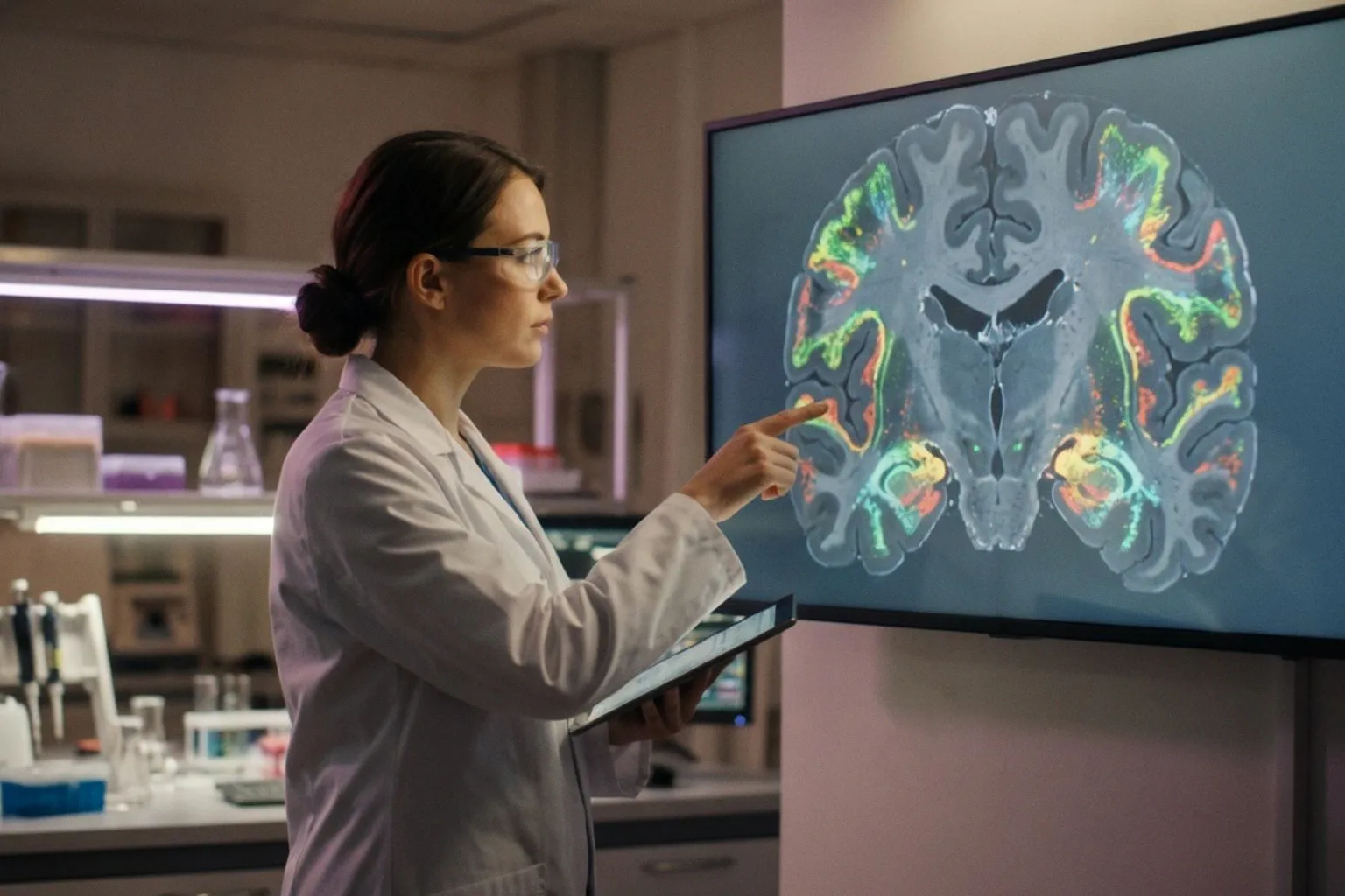
For decades, one of the most persistent frustrations in neuroscience has been the trade-off between knowing what a brain cell is and knowing where it lives. A neuron sitting in cortical layer 5, sending its axon down into the spinal cord, carries a fundamentally different functional identity than a layer 2/3 neuron projecting to other cortical areas — yet the moment tissue is dissociated for single-cell RNA sequencing, that spatial identity is destroyed. Spatial transcriptomics, as detailed in a recent Technology Networks overview of the field, is now closing that gap by mapping gene expression within intact tissue while preserving cellular coordinates, and the implications for brain atlasing and neurodegeneration research are substantial.
Why Position Is Not Optional in the Brain
Consider the sheer organizational density: the human brain contains an estimated 170 billion cells arranged across hundreds of molecularly distinct types, distributed through subregions with markedly different functions — from the six-layered neocortex to the granule cell layers of the cerebellum and the evolutionarily ancient circuits of the brainstem. Within the neocortex alone, each laminar layer houses a characteristic complement of glutamatergic projection neurons and GABAergic interneurons, and those populations project to anatomically distinct targets. A molecular marker expressed in different layers can indicate cells with entirely different circuit roles, which means that standard bulk RNA sequencing — averaging transcriptomic signal across all cells in a tissue homogenate — collapses precisely the diversity we most need to understand. Single-cell approaches recover molecular identity at individual-cell resolution, but the dissociation step erases spatial coordinates: where a profiled neuron resided, which cellular types bordered it, and whether it occupied a plaque-proximal or plaque-distal microenvironment in a diseased brain are all unrecoverable from dissociated data alone.
From Reference Maps to Mechanistic Insight
Early in situ hybridization surveys, including the original Allen Mouse Brain Atlas, demonstrated that brain gene expression is spatially patterned at high resolution, but those approaches were neither genome-wide nor single-cell. The current spatial biology landscape — spanning sequencing-based platforms, imaging-based transcriptomics, and multiplexed spatial proteomics — now provides tools for molecular profiling without that loss of context. Landmark atlas projects are applying these technologies at scale, defining thousands of molecularly distinct cell types and mapping their regional distribution across the brain. This shift allows us to move beyond cataloguing cell types in isolation and toward understanding how gene expression gradients define anatomical region boundaries, how cell-type identity varies across cortical layers, and how disease-associated molecular signatures are spatially constrained to particular circuits or cell populations. For neurodegeneration research specifically, the ability to situate molecular pathology within its precise tissue microenvironment — rather than averaging it away — represents a qualitative change in what we can ask of a brain sample.
What This Means for the Translational Pipeline
The practical relevance for anyone working at the intersection of neuroimaging and clinical neuroscience is considerable. Spatial transcriptomic datasets are beginning to inform how we interpret MRI-derived biomarkers: if a regional signal change on imaging corresponds to a spatially confined molecular signature, the biological grounding of that imaging marker strengthens. Conversely, atlas-level spatial maps can reveal that what appears homogeneous on a scan actually contains functionally distinct subpopulations with different vulnerability profiles. The field is still in an early, atlas-building phase — the reference maps are being drawn — but the trajectory toward integrating spatial molecular data with in vivo imaging is clear. For software developers and methodologists in the neuroimaging space, this convergence signals that future analysis pipelines will need to accommodate multi-modal, spatially resolved molecular layers alongside traditional structural and functional MRI data. The brain, it turns out, has always insisted on being understood in context — and the tools are finally catching up.