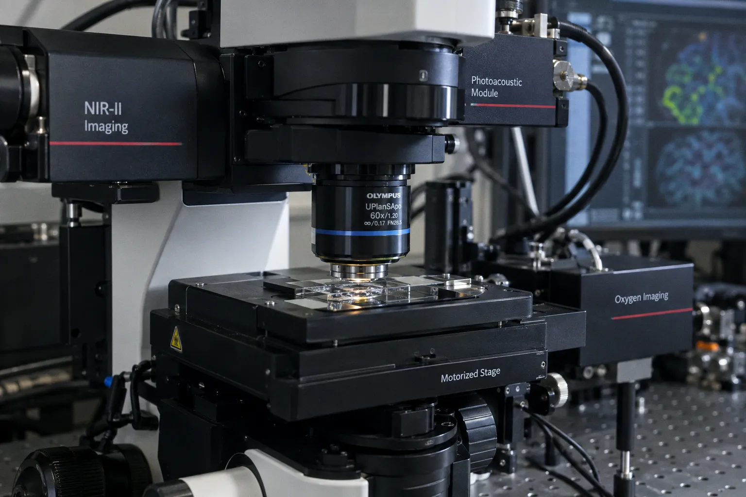
A hybrid microscope from Washington University in St. Louis locks two optical modalities to a single field of view, recording neuronal calcium transients and red blood cell oxygen release in awake mice for the first time at single-cell resolution. According to Medical Xpress, the system — developed by Song Hu's group at the McKelvey School of Engineering and published in Nature Communications — fuses two-photon microscopy with photoacoustic microscopy into a platform the team calls TPM-PAM. The practical payoff is the ability to probe neurovascular coupling directly, rather than inferring it from downstream hemodynamic surrogates.
The Hardware Coupling
Two-photon microscopy tolerates scattering tissue by restricting excitation to a diffraction-limited focal volume, yielding fluorescent calcium signals from individual neurons. Photoacoustic microscopy tolerates the same scattering regime by delivering pulsed laser energy absorbed by hemoglobin; the resulting thermal expansion generates ultrasonic waves that are reconstructed into maps of vessel architecture and oxygen saturation. Each modality alone forces the researcher to choose between reading the neuron or reading the fuel line. The Hu group's contribution is simultaneous acquisition — no sequential scanning, no temporal drift between the two signal channels — at a shared cellular scale.
What the Platform Yields
During whisker stimulation in awake mice, TPM-PAM tracked calcium dynamics in neurons while concurrently resolving how nearby capillaries altered their oxygen delivery. Using tightly focused laser pulses, the team blocked a single capillary or activated a single neuron and observed the resulting perturbations in surrounding tissue and vasculature. The platform moves the experiment from correlative observation toward causal interrogation of how oxygen supply constrains neural computation — and where that constraint fails in stroke and neurodegeneration. Hu, as reported by Medical Xpress, described the work as the first demonstration of simultaneous imaging of neuronal activity and oxygen delivery at the single-cell level.
The Translation Gap
Results remain confined to animal models. TPM-PAM sits at a different point in the imaging hierarchy than clinical fMRI: BOLD fMRI infers neuronal activity from blood oxygenation because it cannot resolve either signal directly. TPM-PAM resolves both, but through optical access that the human skull does not permit without invasive windows. What the technique supplies today is a mechanistic reference standard — ground truth on neurovascular coupling that can inform how BOLD signal models are constrained in software, how acquisition protocols are tuned, and which clinical imaging biomarkers actually track the physiology they claim to represent. The engineering constraints to watch are laser power density, scan depth ceiling, and motion artifact in awake preparations — the same class of bottlenecks that deep-learning reconstruction is currently loosening for structural MRI. Whether the same algorithmic acceleration reaches hybrid optical platforms will determine whether this resolution stays in the lab or enters the translational pipeline.