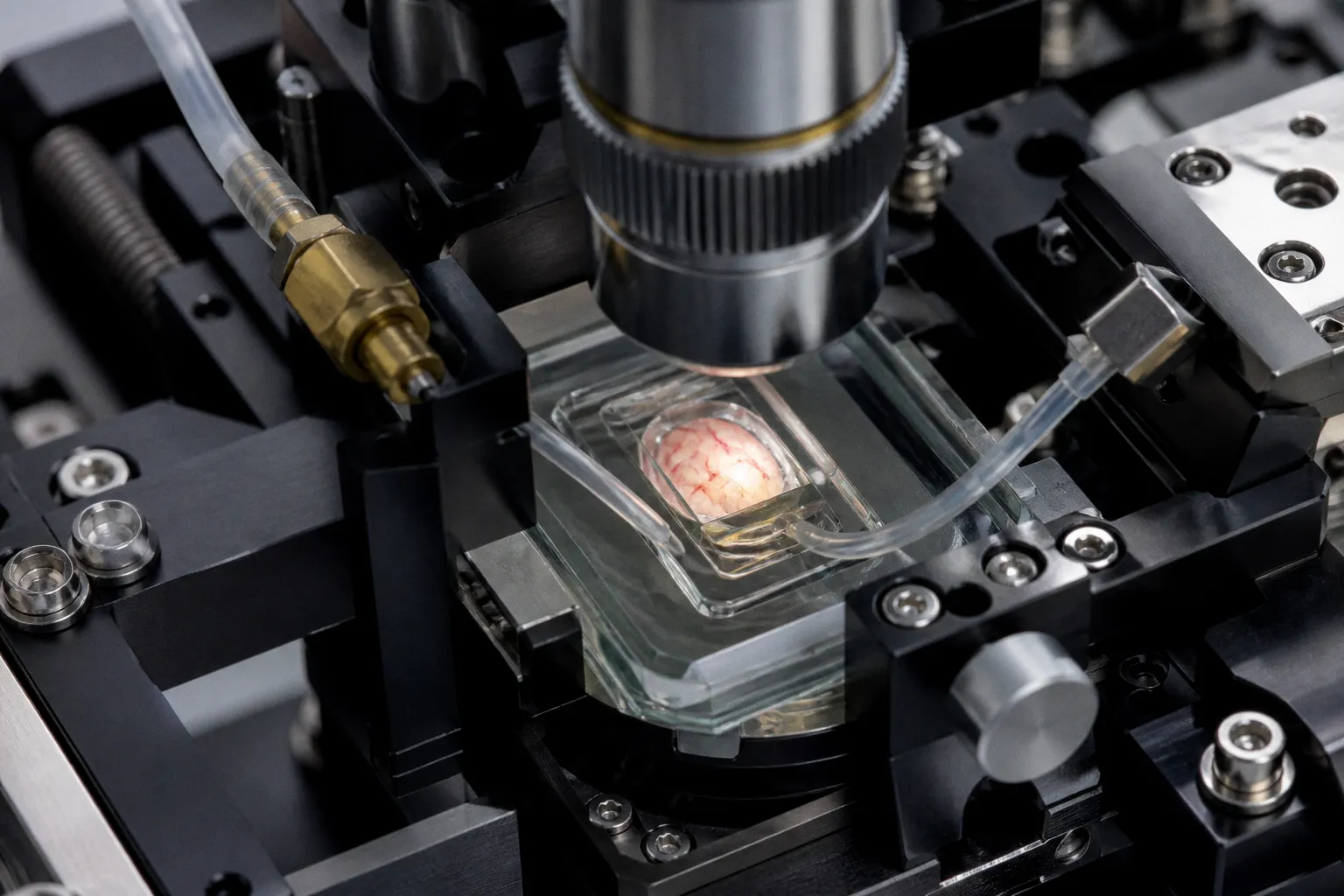
The headline points to a familiar but persistent neuroimaging challenge: vascular structure and tissue oxygenation are related, yet they are not interchangeable measurements. For researchers working at the boundary between MRI software and translational neuroscience, the important question is therefore not whether another brain map is possible, but what additional biological contrast this method may eventually offer—and how carefully that contrast can be validated.
A map with two biological dimensions
Photoacoustic imaging is presented here as a way to capture both vascular anatomy and oxygenation, using two frequencies. The available source material does not provide the imaging protocol, spatial or temporal resolution, acquisition depth, reconstruction method, sample size, or quantitative validation, so the result should be treated as an early technology signal rather than a complete performance claim.
That distinction matters. A vessel map can describe where blood-carrying structures are located, while an oxygenation map may indicate how the local blood environment differs across the imaged tissue. These outputs answer different biological questions, even when they are collected within the same experiment. Consider the implications for analysis pipelines: combining the channels may make regional patterns easier to examine, but it also creates more opportunities for contrast-specific artefacts, calibration problems, or overinterpretation if the relationship between the measurements is assumed rather than tested.
For neuroimaging specialists, the central issue will be whether the dual-frequency acquisition produces reproducible information that remains meaningful across animals, brain regions, and experimental conditions. Without those details, the headline supports interest, but not yet a judgment about sensitivity, specificity, or superiority over established imaging approaches.
Why this matters for translational imaging
The mouse brain is a useful setting for developing imaging methods, but it is also a controlled one. A technique that can map vessels and oxygenation in this context may help researchers study how vascular features and tissue physiology change together over time. However, the trajectory from a preclinical imaging demonstration to clinical neuroimaging is long, and the available evidence does not establish that this approach is ready for human use or for direct comparison with MRI-based oxygenation methods.
This is where software engineering becomes part of the biological question. Any practical assessment would need to examine how the two frequency channels are registered, separated, normalized, and converted into interpretable maps. It would also need to distinguish genuine physiological variation from changes introduced by acquisition geometry, motion, illumination, or reconstruction. None of these technical details is included in the supplied report, so they should remain open checkpoints rather than assumed strengths.
A separate Scientist Live headline reports high-speed microscopy revealing electrical activity across the brain. That is a different imaging direction, but the pairing is instructive: contemporary neurotechnology is increasingly trying to connect structure, physiology, and function rather than treating them as isolated layers. The challenge is maintaining a clear boundary between what an instrument detects and what a biological model infers.
What to watch next
The next useful evidence would be a full methods report describing the dual-frequency design, the animal preparation, validation against an independent measure of vascular or oxygenation status, and the stability of the maps across repeated acquisitions. Quantitative comparisons would be especially important, because a visually compelling overlay does not by itself establish that the two contrasts are accurate or clinically informative.
For now, the restrained reading is the most useful one. Bioengineer.org has identified a potentially valuable way to examine vascular structure and oxygenation together in the mouse brain, but the supplied evidence does not yet show how far the method can travel along the translational pathway. In neuroimaging, that distance is often determined less by the first striking map than by longitudinal reproducibility, transparent reconstruction, and the ability to preserve biological meaning through every stage of analysis.