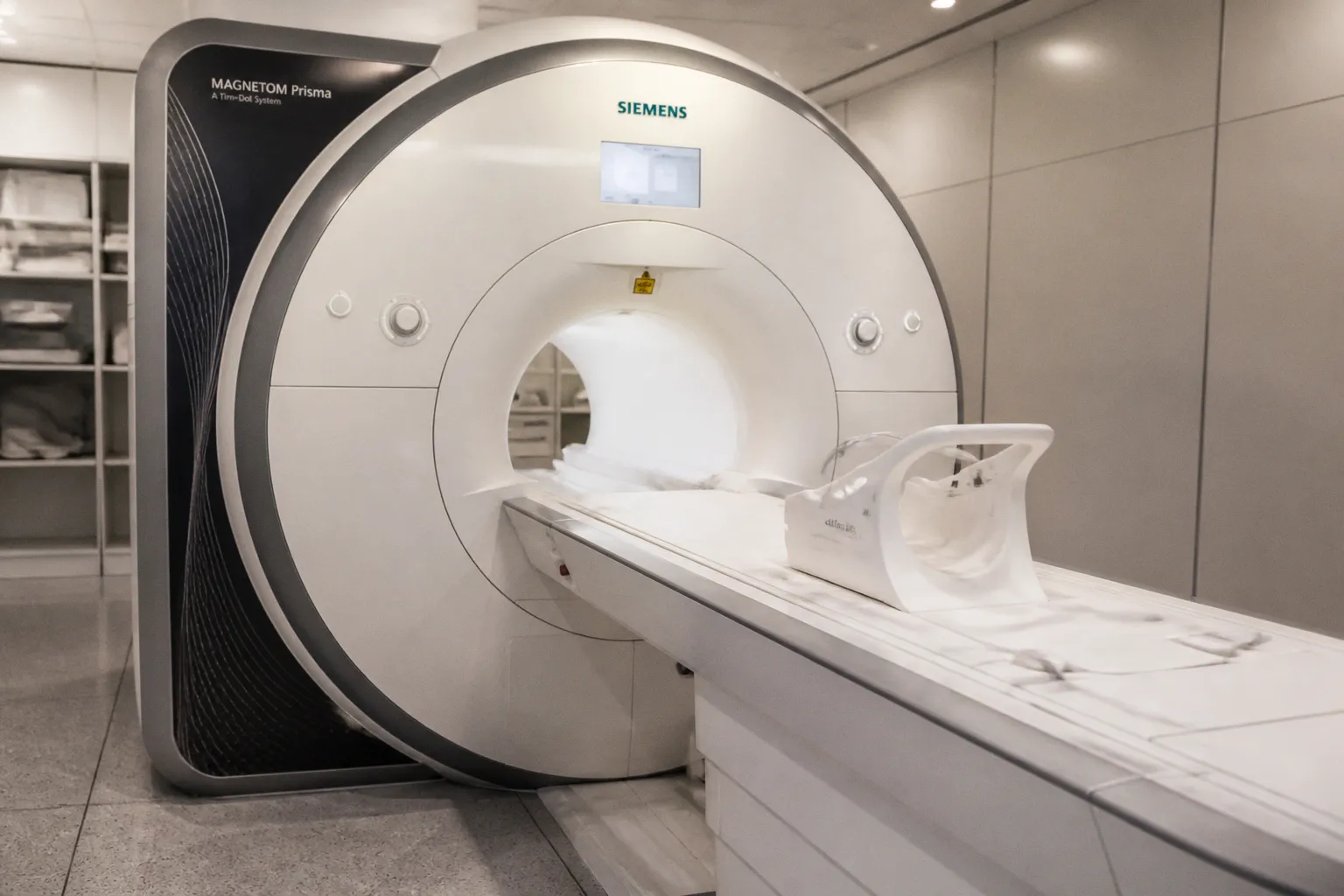
A new review in the Japanese Journal of Radiology, surfaced through JoVE Visualize, frames a quiet but consequential shift in how we read the brain and spinal cord with MRI — away from visually inspected images and toward truly quantitative measurements. The survey covers four territories that matter to anyone working at the interface of imaging software and clinical interpretation: deep-learning reconstruction and acceleration, quantitative oxygen-metabolism assessment, standardized spinal-cord imaging, and diffusion-based monitoring of the glymphatic system. This shift allows us, for the first time in routine practice, to treat the scan less as a picture to be inspected and more as a dataset to be measured.
From pixel to physiology
Consider what this transition means at the console. Deep-learning reconstruction is not simply making prettier pictures faster; it is recovering tissue contrast that earlier pipelines quietly smoothed away, the kind of contrast that, tracked along a longitudinal trajectory, distinguishes genuine degradation from noise or age-related drift. Oxygen-metabolism quantification pushes past the structural envelope into the metabolic substrate itself, giving us a window onto how neurons use energy at rest, during a task, or in the earliest phases of disease. Perhaps most provocatively, diffusion MRI aimed at the glymphatic system offers a non-invasive look at how the brain clears its own waste — a process long implicated in neurodegeneration but rarely visible to the clinician's eye. Taken together, these threads point toward imaging protocols whose outputs can be compared across sites, across scanners, and across years without the usual anxieties about reconstruction drift.
The spinal cord comes into focus
If the brain has long dominated the neuroimaging conversation, the spinal cord is where this cluster of publications lands some of its most practical weight. A study in Biomedical Signal Processing and Control describes a 3D deep-learning model that reconstructs higher-resolution spinal-cord images from the lower-resolution scans most clinical centers realistically acquire, explicitly to improve segmentation and volumetric assessment. The method is applied to longitudinal multiple-sclerosis atrophy measurement and therapy monitoring — use cases where fractions of a millimeter of cord diameter, watched across months, can reshape how we counsel a patient about disease trajectory. Reporting on a parallel line of work, Multiple Sclerosis News Today highlights MRI maps that reveal distinct brain network patterns in people with MS, reinforcing the broader message that quantitative imaging rewards patience: less about a single snapshot, more about reading patterns across time.
Adjacent territory worth watching
Slightly outside the MRI orbit but worth a brief note, a TipRanks report covers Gestala's RMB 570 million raise to accelerate an ultrasound-based brain-computer-interface and brain-AI platform. The modality is different, yet the underlying ambition — treating brain–machine interaction as a quantitative, data-rich discipline rather than a one-off demonstration — runs parallel to the radiology review's vision for MRI. For engineers and researchers building pipelines around brain data, both movements point toward the same horizon: reproducible, comparable numbers where there used to be only images.