The technique, designated DEPICTA, runs as a deep learning-reconstructed multi-contrast multi-shot EPI acquisition and held clinical image quality across T1-FLAIR, T2-FLAIR, and DWI in the evaluated cohort.
Acquisition and reconstruction pipeline
DEPICTA attacks the latency bottleneck at the reconstruction stage. Multi-shot EPI partitions k-space into individually navigable segments, which constrains motion-induced phase errors that would otherwise alias into the final image. The deep learning model then resolves the inverse problem from undersampled data — a regime where conventional parallel imaging tolerates only modest acceleration factors before SNR degrades past diagnostic thresholds. The reported preservation of signal-to-noise ratio and clinical image quality across all three contrast families suggests the network did not memorize a single contrast signature. The reduction in motion artifacts among uncooperative patients is a direct consequence of shorter acquisition windows: when the scan terminates before bulk head motion accumulates, ghosting and intra-volume blurring collapse on their own.
A parallel hardware lever
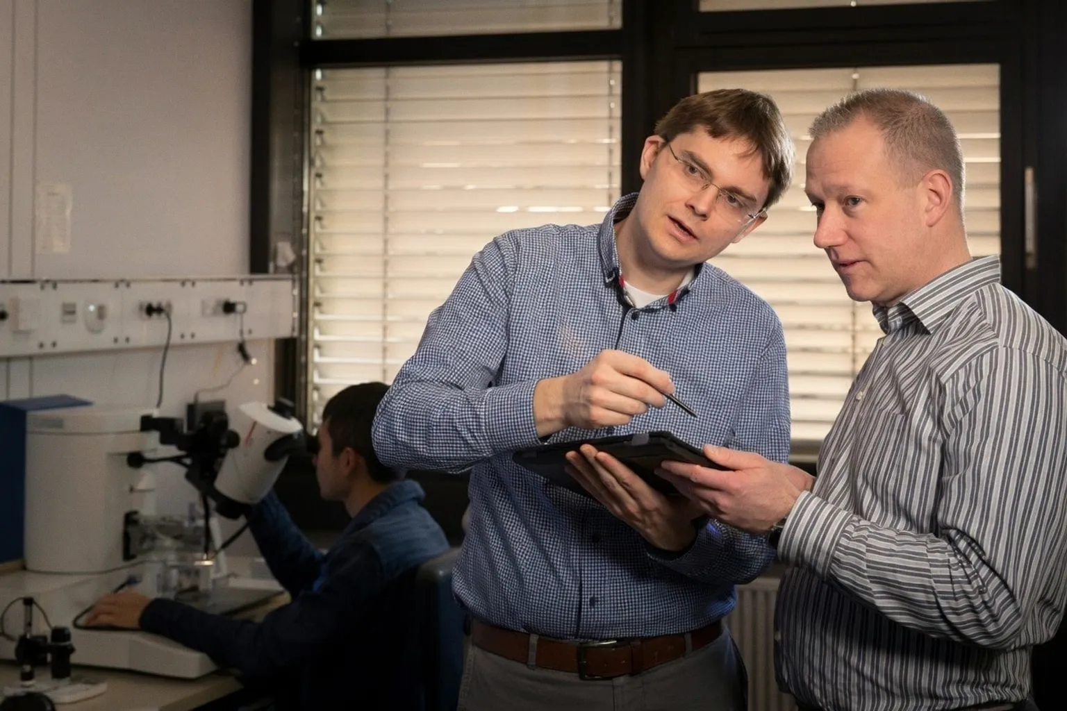
The 100-second ceiling is not held by reconstruction alone. A separate line of work in Science Advances, led by Prof. Jens Anders and Michal Kern at the University of Stuttgart's Institute of Smart Sensors, attacks the receiver dead time — the blind interval after an RF excitation pulse during which the resonant circuit's stored energy drowns the weak spin signal. According to the team, shifting the excitation energy to a different frequency allows simultaneous excitation and detection, yielding recovery orders of magnitude faster than the conventional wait-and-decay path and recovering ultrafast-decaying signals that standard systems forfeit. The group is collaborating with Ulm University Hospital on clinical translation, and with Charité Berlin and the Helmholtz-Zentrum Berlin on electron paramagnetic resonance, where signal lifetimes are orders of magnitude shorter still and where the same frequency-shifting principle is being tested for skin cancer imaging.
Where this leaves the scanner
Two distinct levers, one ceiling. Reconstruction algorithms compress the time required to fill k-space to a diagnostic criterion; hardware physics extends the detectable signal lifetime at the receive coil. For software developers building deep learning reconstruction pipelines, the DEPICTA result establishes a clinically validated floor — under 100 seconds with preserved diagnostic content across multiple contrasts — and removes one of the standard objections to accelerated brain protocols. For scanner engineering, the Stuttgart result exposes a different constraint: the dead time that gates access to short-T2 tissue compartments, with a frequency-shifting workaround already demonstrated. The two efforts converge on the same practical outcome: shorter, quieter acquisitions without diagnostic compromise.