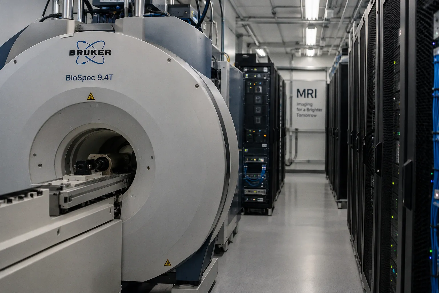
The optical barrier, cleared
According to News-Medical, Bruker has pushed whole-organism 3D imaging forward through enhanced tissue-clearing workflows — a methodological advance that addresses photon scattering at the sample-preparation stage rather than compensating for it during reconstruction.
The headline is sparse. The physics underneath is denser. Intact biological tissue scatters excitation photons through refractive index mismatch between lipid, protein, and aqueous compartments — a hard depth ceiling for conventional confocal and light-sheet microscopy. Tissue-clearing protocols — solvent-based or aqueous — homogenize those indices across the specimen, extending useful imaging depth into regimes where whole-organ and whole-organism volumes become tractable.
For MR-focused workflows, the constraint is direct: cleared tissue cannot yield standard T1 or T2 contrast because the chemical environment generating MR signal has been altered. What clearing delivers is anatomical and connectomic mapping at mesoscopic scale, not diagnostic MR contrast. The two modalities occupy adjacent, not overlapping, territory — and that boundary defines where the announcement matters to this readership.
Adjacent signal, same pipeline
Two parallel developments bracket the practical context. A medRxiv preprint evaluates deep learning-based reconstruction against automated brain segmentation in accelerated T2-weighted MRI, probing volumetric robustness as undersampling ratios increase. OpenNeuro has released the GRAND repository — multi-modal MRI and fMRI across 109 older adults — as a benchmark surface for cognitive aging algorithms.
Together with tissue-cleared volumes, these inputs form a coherent data pipeline: ground-truth connectomics upstream, reconstruction-aware segmentation in the middle, open validation cohorts downstream. Each layer constrains the next. A workflow that lowers the resolution floor for cleared-tissue imaging raises the demand on segmentation algorithms to match that resolution without hallucinating boundaries in undersampled MR acquisitions — which is precisely the failure mode the medRxiv study is designed to measure, and precisely the kind of failure an open cohort like GRAND can stress-test at scale.
What requires verification
The Bruker announcement carries no published protocol in the available record. The "enhanced" qualifier demands specifics: clearing chemistry, refractive index target, acquisition modality, resolution floor, acquisition time per specimen. Without those parameters, performance against established protocols — uDISCO, iDISCO, CLARITY — cannot be assessed on signal-to-noise or throughput grounds.
For the neuroimaging software audience, the relevant trade-off is sharper still. Cleared-tissue workflows degrade the chemical contrast MR relies on; deep-learning reconstruction tolerates acceleration but constrains segmentation accuracy; open datasets calibrate but do not validate. Until the Bruker workflow parameters surface — refractive index target, scanner-agnostic vs. modality-bound acquisition, compatibility with downstream segmentation pipelines — the announcement remains a directional signal, not a benchmark.