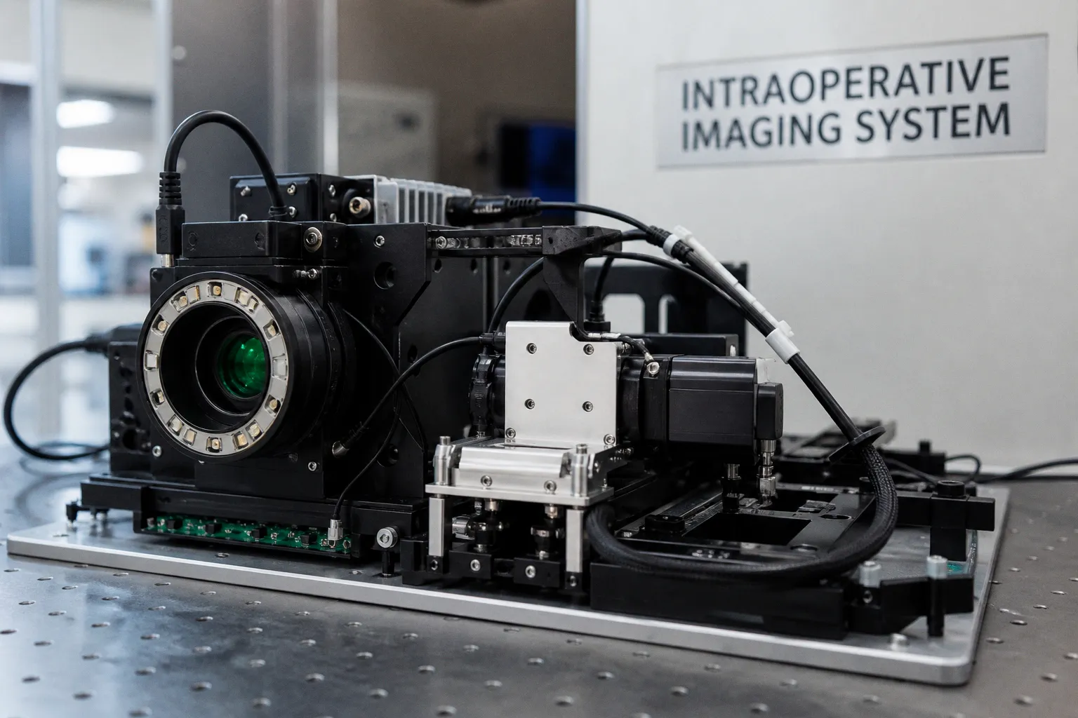
A neurosurgeon peels back the dura, navigates to a glioma infiltrating eloquent cortex, and must decide in real time how much tissue to resect without devastating the patient's speech or motor function — a choice that has always balanced anatomical landmarks against intraoperative judgment. A new perspective published in npj Biomedical Innovations reviews the integrated platforms now attempting to tip that balance by fusing structural, functional, molecular, and optical imaging modalities into unified surgical workflows, assessing AI-driven multimodal fusion as genuinely promising yet still constrained by early-stage clinical evidence and the absence of large-scale validation. For anyone working at the boundary between neuroimaging software and the operating room, the review offers both a candid catalogue of what is available and a sober reminder that the translational pipeline remains incomplete.
What the Perspective Actually Covers
The review maps an expanding landscape: structural MRI sequences that define tumor margins, functional data that localize language and motor networks, molecular imaging that characterizes tissue at the biochemical level, and optical techniques that provide real-time contrast during resection. Consider the implications of bringing these streams together onto a single platform rather than toggling between separate viewers during surgical planning. A unified dashboard promises to reduce the cognitive load on surgeons who currently mentally register functional maps against anatomical volumes, a process prone to subtle degradation under time pressure. The perspective argues that this integration is not merely convenient but potentially transformative for personalized brain tumor care, aligning surgical strategy with the individual trajectory of each patient's disease.
AI-Driven Fusion: Promise Tempered by Evidence
The paper identifies AI — particularly deep-learning segmentation and multimodal registration algorithms — as the engine that could make real-time fusion practical at scale, yet it is careful to frame this optimism within clinical reality. Current evidence, the authors note, remains early: most validation studies involve small cohorts, retrospective designs, or single-institution protocols, and insufficient large-scale prospective trials mean that generalizability is still an open question. This is a familiar pattern in translational neuroimaging; algorithmic performance in controlled datasets does not automatically survive the variability of the scanner-to-scalpel continuum. For software developers and radiologists evaluating these platforms, the key takeaway is that regulatory clearance and publication in a conference proceeding are not substitutes for longitudinal outcome data linking the tool to measurable improvement in extent of resection or progression-free survival.
Where This Leaves the Field
Brain tumors remain among the most challenging malignancies, and the perspective frames multimodal AI integration not as a solved problem but as a trajectory — one that requires standardized protocols, interoperable data formats, and multicenter validation before it can reliably enter clinical decision-making. The shift from siloed imaging sequences to unified, AI-augmented platforms allows us to imagine surgical planning that adapts in real time to intraoperative findings, but realizing that vision depends on engineering pipelines robust enough to handle heterogeneous data without introducing artifacts or latency. For clinicians, the near-term question is which components of multimodal fusion are mature enough to incorporate into current workflows and which deserve continued caution. For researchers and developers, the perspective underscores a persistent gap: the field has sophisticated tools and compelling proof-of-concept results, yet the bridge to large-scale, reproducible clinical impact is still under construction.