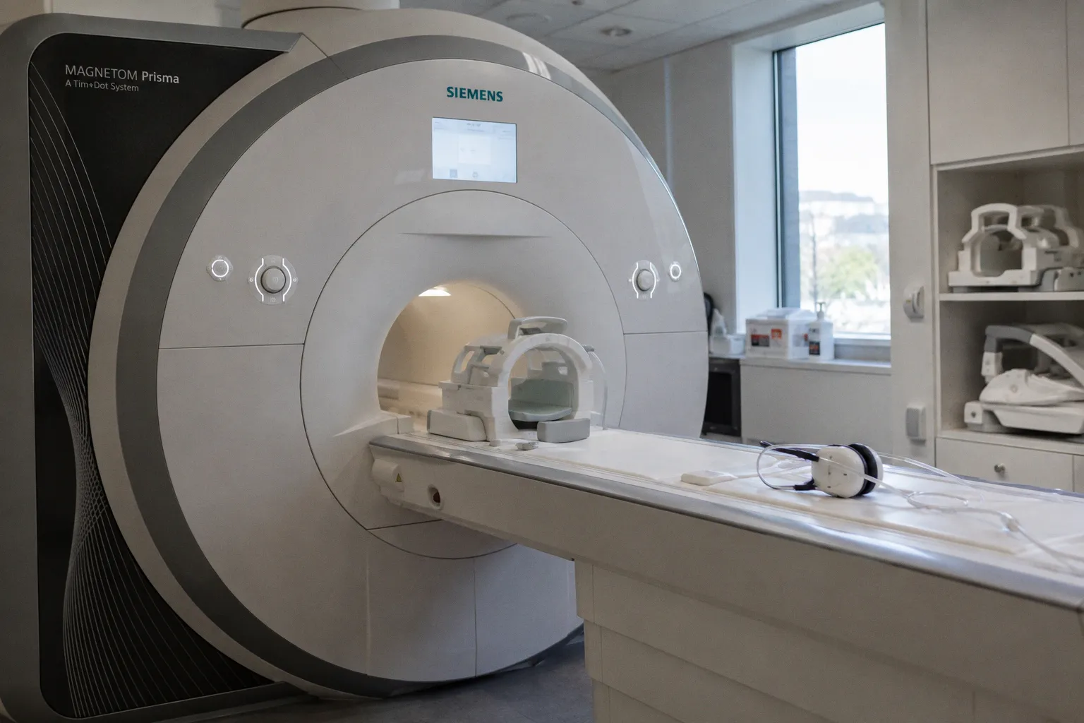
When a patient with Graves' disease begins losing vision, the clinical question is straightforward — is the optic nerve being compressed, and can we intervene before irreversible damage sets in? What is far less obvious, and what a new study in Frontiers in Neuroscience begins to address, is whether the brain itself has already begun to reorganize in ways that complicate recovery. Researchers used resting-state functional MRI to examine disrupted functional connectivity patterns in patients with dysthyroid optic neuropathy, identifying network-level biomarkers that appear to correlate with the degree of clinical visual impairment — a finding that nudges us closer to understanding how chronic orbital disease reshapes cortical architecture over time.
Mapping Connectivity Beyond the Optic Nerve
Dysthyroid optic neuropathy, the vision-threatening endpoint of thyroid eye disease, has long been understood through the lens of mechanical compression: inflamed extraocular muscles thicken within the confined orbital space, and the optic nerve pays the price. This study, however, looks past the orbit and into the brain itself. The researchers evaluated resting-state functional MRI metrics — specifically degree centrality and voxel-mirrored homotopic connectivity — to detect alterations in how distributed brain networks communicate when the visual system has been chronically compromised. Degree centrality, which reflects the number and strength of functional connections a given brain region maintains with the rest of the network, offers a window into whether visual cortex hubs are losing their integrative role. Voxel-mirrored homotopic connectivity, meanwhile, measures the synchrony between homologous regions in the two hemispheres; a reduction here can signal that bilateral coordination — essential for coherent visual processing — has begun to fray. Consider the implications: if these metrics reliably track with the severity of a patient's visual deficits, they become candidate biomarkers not just for diagnosis but for longitudinal monitoring of treatment response.
Why Resting-State Protocols Matter Here
What makes this work particularly relevant for neuroimaging practitioners is the reliance on resting-state paradigms rather than task-based designs. Patients with significant visual impairment cannot easily perform stimulus-driven tasks inside the scanner — a practical constraint that makes resting-state acquisition not merely a convenience but a methodological necessity. This shift allows us to assess cortical reorganization without demanding visual compliance from the very population whose visual function is compromised. For those building or refining analysis pipelines, it also underscores the importance of robust preprocessing: head motion, physiological noise, and signal-to-noise considerations become especially critical when the expected connectivity differences may be subtle degradation patterns layered on top of normal variability. The choice of degree centrality and homotopic connectivity as analytical frameworks reflects a growing preference in the field for graph-theory-informed metrics that capture network topology rather than isolated regional activation — an approach that aligns well with the broader trajectory of connectomics research.
A Broader Context and What to Watch
This study arrives within a wider moment for functional connectivity research. Independent work recently highlighted in Nature has explored how resting-state brain age estimates relate to cognitive and sensorimotor abnormalities in schizophrenia spectrum disorders, reinforcing the principle that resting-state fMRI can reveal clinically meaningful signatures across very different patient populations. Meanwhile, efforts to complete the connectome of the Drosophila brain, reported by Phys.org, remind us that mapping connectivity — whether in a fruit fly or a human thyroid patient — remains one of neuroscience's most persistent and productive undertakings. For the neuroimaging community, the key question going forward is whether the aberrant connectivity patterns identified in dysthyroid optic neuropathy prove reversible with successful decompression or immunosuppressive therapy, or whether they reflect a more permanent cortical remodelling that sets a ceiling on visual recovery. Longitudinal follow-up with serial fMRI will be essential to resolve this, and it is precisely the kind of translational gap that software developers and clinicians working together can begin to close.