A new framework from Nature Neuroscience invites clinicians and imaging scientists to reconsider something many of us take as a given: that the brain's hierarchy of information flow is a fixed scaffolding on which cognition hangs. As reported in the study, which pairs integrated effective connectivity with an unconstrained signal-flow mapping technique, cortical hierarchies do not sit still — they reorganize depending on whether a person is turned outward toward the world or inward toward their own internal state.
From laminar structure to living dynamics
The team's approach, built around what they term integrated effective connectivity (iEC), recovers the directionality of connections within the human connectome and aligns them with the feedforward and feedback pathways that histologists have long described across cortical layers. What makes this more than a methodological exercise is the trajectory it traces: a monotonic gradient running from sensorimotor regions through association cortex and onward into paralimbic territory, echoing the structural model of laminar connectivity that has quietly guided cortical neuroscience for years.
The clinical relevance sits in what happens when the brain switches tasks. The hierarchy flattens during externally oriented processing — when attention is anchored to sensory cues — and steepens during internally focused conditions, when interoceptive regions appear to pull more of the signal flow upward. For anyone designing fMRI paradigms or interpreting task-based connectivity, this is a useful reminder that the "default" architecture is itself a moving target, and that the shape of effective connectivity at any given moment reflects the cognitive state being measured.
Resolution matters at the small scale
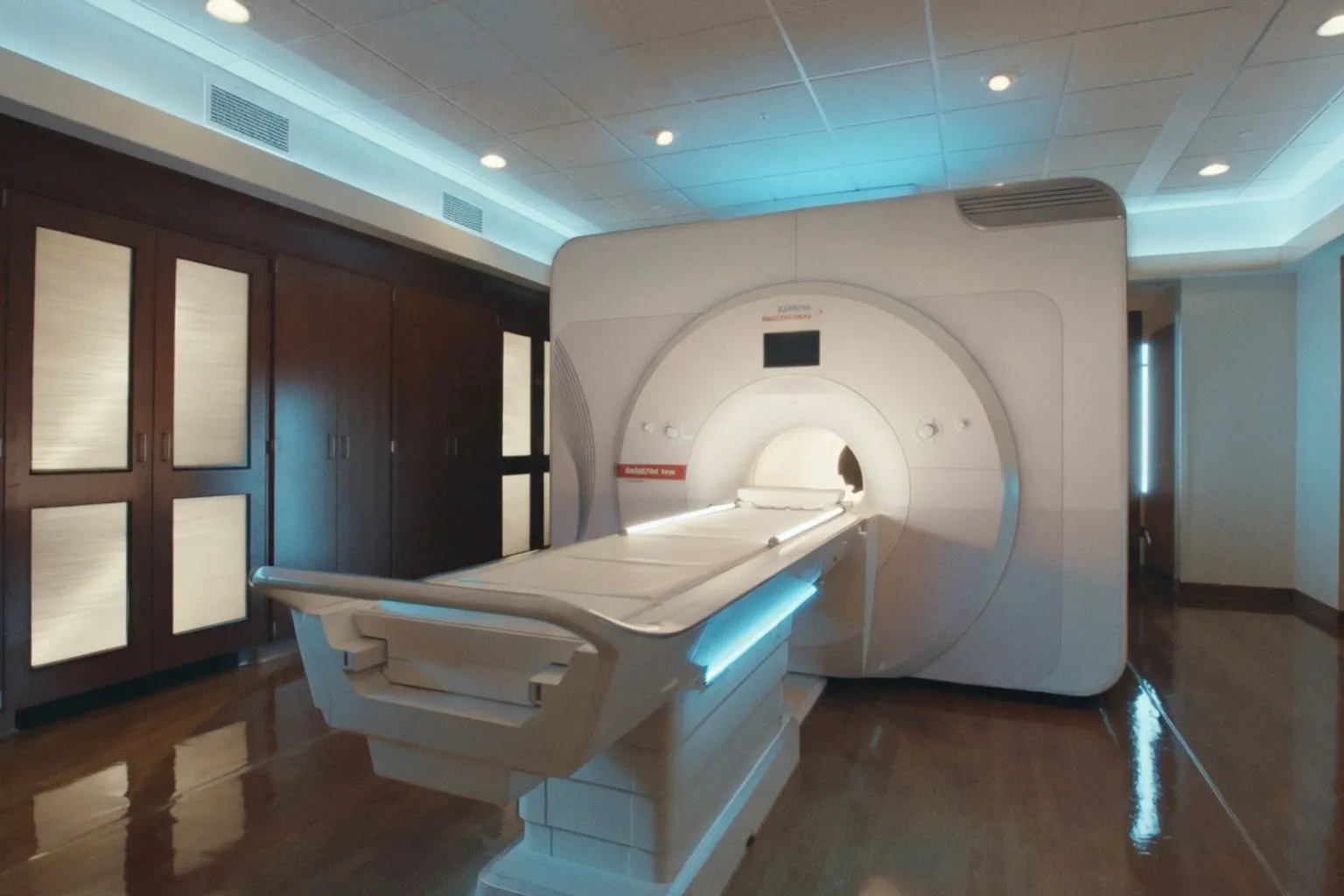
That principle lands with particular force at the Beckman Institute, where researchers have been turning ultrahigh-field MRI toward decision-making circuitry that operates below the threshold of conventional imaging. In their account, standard human brain imaging at 2 to 3 millimeters simply cannot resolve the function of very small structures, which has left regions like the locus coeruleus — a brainstem site only a few millimeters across with an outsized role in arousal and choice — something of a blind spot in routine protocols. The team's 7 Tesla work, developed through the Carle Illinois Advanced Imaging Center as a collaboration between the University of Illinois Urbana-Champaign and Carle Health, pairs submillimeter resolution with a gambling-style task that includes high-stakes trials designed to elicit stronger, more detectable neural responses. This shift allows us to trace decision processes through pathways that were previously inferred rather than imaged directly.
Where connectomes meet cognition
A complementary thread is emerging from work published in eLife, where researchers used deep learning alongside fMRI and PET data to predict memory performance from functional connectomes across both resting-state and task conditions. The study introduces a "brain cognition gap" measure that appears to track physical activity, cardiovascular risk, dopamine binding, and broader cognitive resilience — a quietly ambitious effort to compress an individual's functional architecture and behavioral profile into a single, clinically legible quantity.
Consider the implications for your own pipelines. Effective connectivity analyses, once dominated by assumptions of stationarity, are now being asked to accommodate state-dependent reorganization rather than average it away. Acquisition strategies, particularly at ultrahigh field, are yielding usable signal from structures that sat below the resolution floor for most of the imaging era. And machine learning approaches are beginning to quantify not just what a connectome looks like, but how far it has drifted from the cognitive performance it ought to support. The throughline is subtle but worth holding onto: the brain we map is not the brain we measure a moment later, and the tools we build now are finally catching up to that fact.