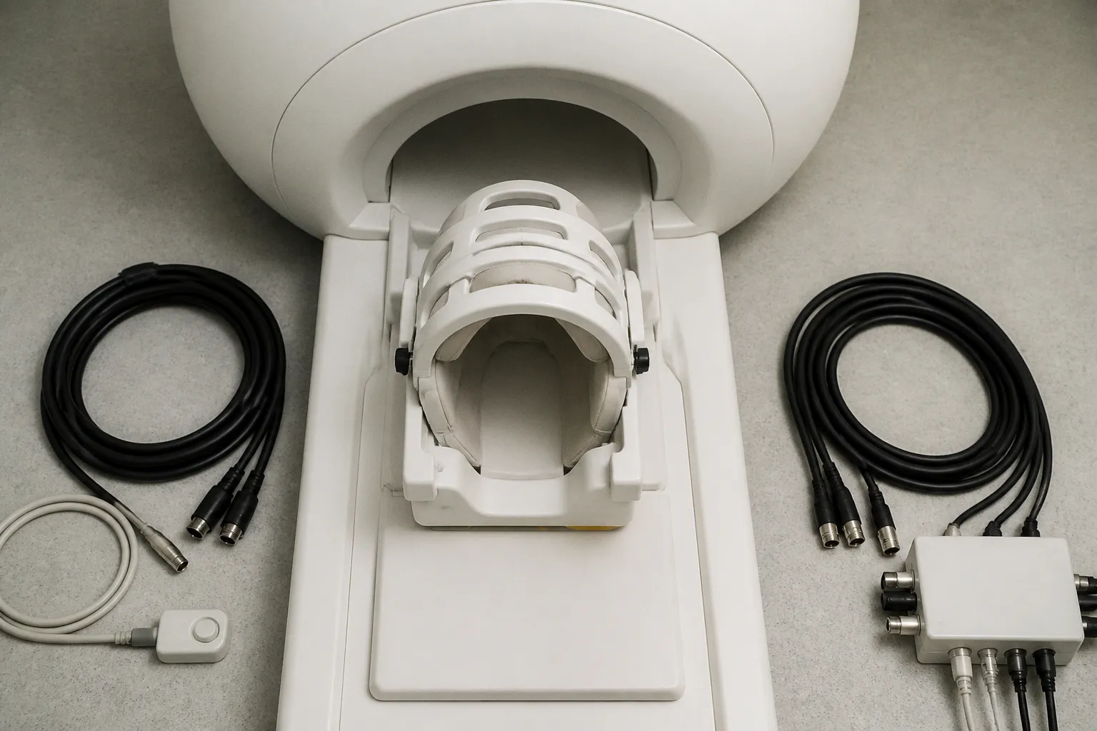
The spatial scale that bridges single neurons and whole-brain networks has long sat in a quiet corner of human neuroscience, starved of tools that could reach it. According to a recent essay collection in The Transmitter, that is beginning to change as custom software packages, web-based applications, and video tutorials quietly lower the barrier to functional MRI and high-resolution cortical layer imaging. For clinicians, software developers, and imaging researchers who have watched fMRI pipelines grow ever more demanding, this is a meaningful shift in who can sit at the table — and what questions they can reasonably ask of their data.
When the barrier stops being code
Consider the implications for a young lab that wants to probe columnar or laminar activity but inherits a scanner, a budget, and almost no in-house programming depth. The Transmitter's contributors argue, with some care, that the bottleneck is no longer purely physics — it is workflow literacy. New open toolkits and guided tutorials now walk researchers through acquisition strategies, preprocessing decisions, and layer-fMRI analysis choices that once required months of bespoke scripting. This shift allows us to redirect scarce clinical and engineering time toward interpretation and validation rather than toward reconstructing someone else's pipeline from a forum thread at midnight.
Bridging the spatial gap neuroscience forgot
Human neuroscience has, by the contributors' account, largely overlooked the mesoscopic scale — the territory between cells and brain areas where cortical layers, columns, and microcircuits actually do their work. New advances in functional MRI technology are beginning to make that scale tractable, and emerging methods now permit researchers to combine tactics from opposite ends of the analytic spectrum, what the collection describes as the ability to have one's cake and eat it too: data-driven discovery alongside hypothesis-driven modeling. A better understanding of the blood oxygen level dependent, or BOLD, signal, the authors note, will require more support for multimodal imaging studies that anchor hemodynamics to electrophysiology rather than trusting either in isolation.
Where it is worth keeping watch
For our readers building or buying imaging software, the practical questions are converging quickly. Smaller studies remain essential for testing new scanning paradigms, even as the field debates whether every brain-behavior association study must run into the thousands of participants — a tension that has real consequences for how we design pipelines, allocate compute, and price software support contracts. The Transmitter also places this technical moment inside a broader federal-funding landscape that researchers describe as tumultuous, which means the tools we adopt today will likely have to outlive the grants that paid for them. Worth tracking in the coming quarters: which open packages reach clinical-grade documentation, and which tutorial ecosystems attract the kind of community that survives a funding cycle.