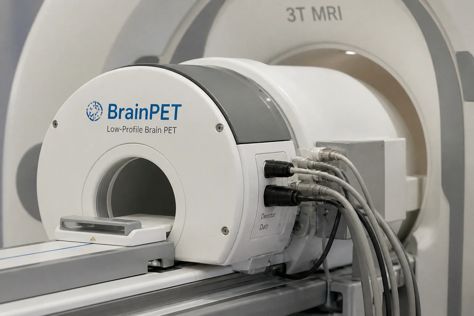
The significance for neuroimaging is not simply that these methods produce more data, but that they may help connect cellular and molecular relationships with the pathology seen in conditions such as Alzheimer’s disease and stroke. A separate development reported by The National Law Review points in a complementary direction: Cubresa has received FDA 510(k) clearance for a BrainPET scanner designed to operate within clinical MRI systems, enabling simultaneous PET/MRI brain imaging.
From anatomical contrast to biological relationships
For clinicians and imaging scientists, the central challenge in advanced brain mapping is rarely the absence of images. It is the difficulty of translating spatial patterns into biology: determining which cellular populations occupy a region, how molecular processes are distributed through tissue, and whether a visible abnormality reflects an active disease mechanism or a later consequence of injury.
The review highlighted by News-Medical.net places single-cell multi-omics and high-resolution imaging within that broader effort. Single-cell approaches can describe molecular features at cellular resolution, while high-resolution imaging preserves the spatial organization in which those cells exist. Consider the implications for diseases with complex and changing tissue environments. A map that links molecular identity to anatomical location may offer a more informative trajectory than either a conventional image or a molecular measurement considered in isolation.
That does not make the resulting maps automatically clinical biomarkers. The confirmed reporting does not establish that these technologies are ready to guide routine diagnosis, predict an individual patient’s outcome, or replace established imaging protocols. Their immediate importance is methodological: they offer ways to examine the relationship between structure, cellular composition, and molecular activity with greater specificity.
Why simultaneous PET/MRI matters to the software layer
The Cubresa announcement concerns a different part of the same translational pathway. According to The National Law Review, the company’s BrainPET scanner received FDA 510(k) clearance and is designed as a low-profile molecular imaging system that operates inside clinical MRI scanners. The reported result is simultaneous PET/MRI brain imaging for clinical applications.
PET and MRI provide different kinds of information. In this context, MRI supplies anatomical imaging while PET contributes molecular imaging, allowing both streams to be acquired together rather than treated as entirely separate examinations. For imaging informatics teams, that combination raises practical questions that are at least as important as the hardware itself: how the datasets are registered, how motion and timing are handled, how image quality is assessed, and how software presents complementary signals without encouraging premature biological conclusions.
The clearance is therefore relevant to more than scanner procurement. It may influence how institutions think about integrating molecular and anatomical imaging within existing MRI infrastructure. At the same time, the available evidence does not provide a detailed account of clinical performance, implementation requirements, workflow effects, or comparative outcomes. Those questions remain part of the evaluation burden for any site considering a new imaging configuration.
The next step is validation, not acceleration
Taken together, the two developments describe a field moving toward richer spatial integration. One line of work seeks to connect high-resolution images with single-cell and molecular relationships; another brings PET-based molecular imaging into an MRI environment. Both are valuable because they address the same underlying limitation: anatomy alone may not explain the full biological state of diseased brain tissue.
The practical lesson is to preserve the distinction between mapping capability and clinical certainty. A more detailed image can expose subtle biological organization, but it can also create new demands for harmonization, interpretation, and longitudinal validation. Before these methods reshape routine brain mapping, researchers and clinical teams will need to establish how consistently the signals can be measured and how reliably they correspond to meaningful disease processes.
For now, the review and the BrainPET clearance mark important pieces of infrastructure for translational neuroimaging. The durable advance will come when those pieces can be connected—spatially, computationally, and clinically—without allowing technical resolution to outrun the evidence needed to interpret it.