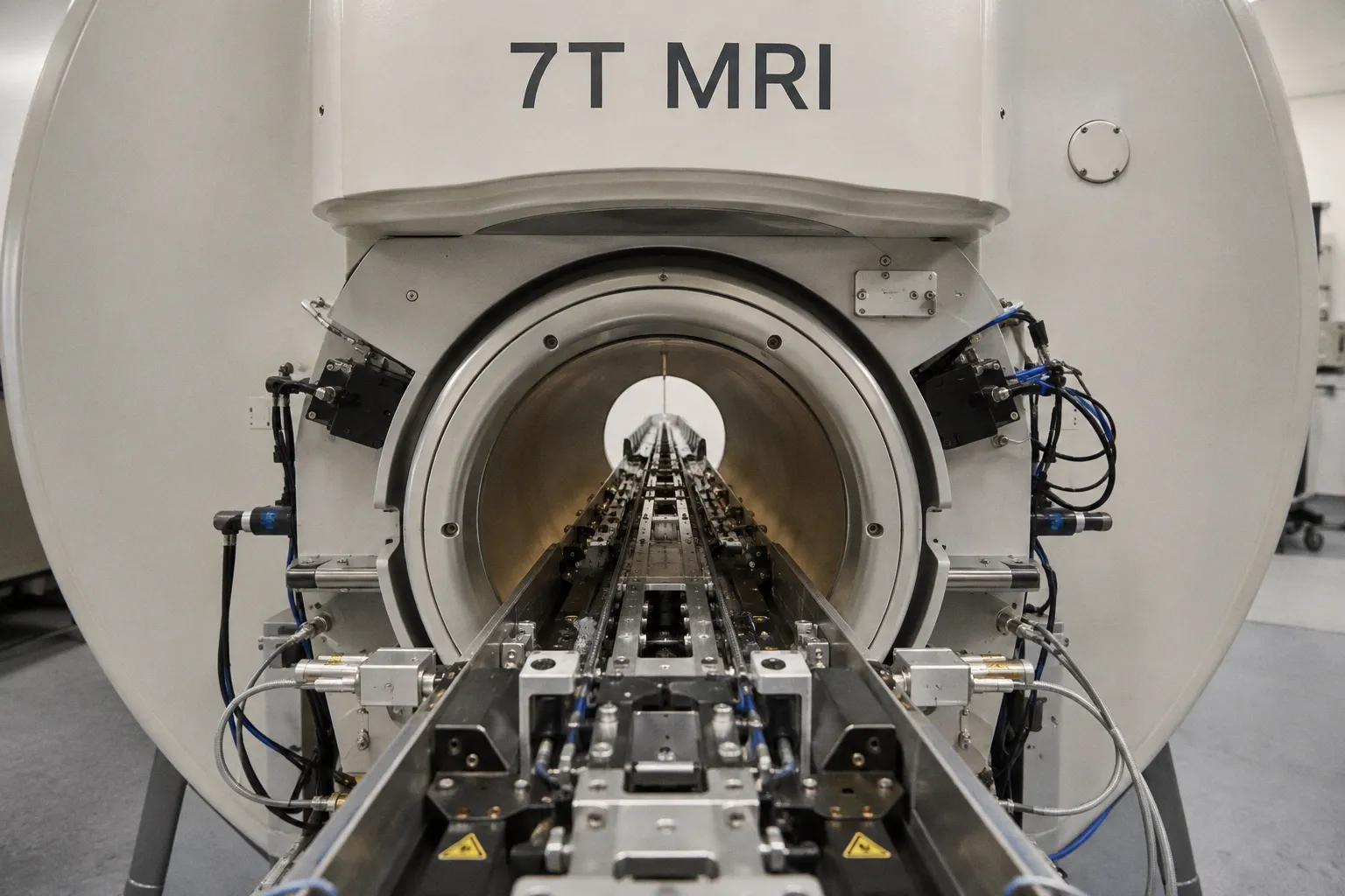
The dataset is now published in Cell, and it sits beside the female connectome the same group completed two years ago. For anyone who treats neuroscience as a signal-reconstruction problem, the resolution and the consistency constraints are the actual news.
Imaging the connectome
The male brain was physically sectioned into 66 slabs, then run through repeated rounds of high-resolution scanning, with each slab contributing a stack of image volumes that were later registered into one 3D map of the brain and ventral nerve cord. Prof Gregory Jefferis of the MRC Laboratory of Molecular Biology, co-lead on the project, described the result as a new telescope for the field. From a pure acquisition standpoint, the bottleneck was not raw voxel size but stitching: 66 independent volumes, each with its own contrast drift and alignment error, had to converge on a common coordinate system tolerant enough to resolve individual axons. The pipeline tolerates that error only because the male and female datasets were reconstructed with the same tooling, which is what makes the comparison statistically usable.
What 5% yields
Roughly 95% of neuronal cell types are shared between male and female fly brains. The remaining ~5% — a small absolute number — drives three male-biased behaviours the authors were able to localize to specific circuits. Visual-tracking wiring that lets a male follow a moving female is enlarged relative to the female template. Aggression-associated circuitry is substantially denser, consistent with the higher frequency of male–male combat. A third, male-unique pathway produces the wing-vibration song used in courtship; if the circuit is mis-tuned, Dr Philipp Schlegel noted, the rejection from an unreceptive female is not subtle. Joint first author Dr Isabella Beckett framed the comparison as confirmation rather than surprise — the headline is the precision of the localization, not the existence of the difference.
Translation, and what to watch
Jefferis is explicit that the dataset does not explain human sex differences. The translational angle is narrower and more useful: the same genetic machinery that reshapes a fly circuit — the two key genes the authors trace here — is implicated in human conditions where wiring goes wrong, including autism and schizophrenia. For a clinical audience, the relevant signal is methodological. Connectomes at this resolution require imaging pipelines that behave identically across years of acquisition, and reconstruction software that can hold 124 million edges without silent merge errors. That engineering bar is what the field has just cleared, and it is what subsequent human-scale efforts will inherit.
For MRI-adjacent workflows, the implication is indirect but real. The fly connectome standardizes how a community validates dense, anisotropic neural reconstructions against ground truth. Any software pipeline that ingests diffusion or structural MRI and outputs tractography at comparable density will now be benchmarked against an external, peer-reviewed whole-brain standard rather than internal consistency alone.