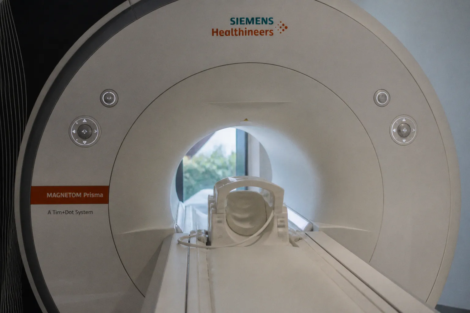
News-Medical reports a long-term MRI finding that sharpens a difficult clinical question: structural brain changes appeared up to seven years before the first signs of amyloid plaque buildup, suggesting that MRI might offer a potential early biomarker for Alzheimer’s disease. The study followed people with regular MRI scans over nearly two decades, giving the reported sequence of events a much longer time frame than a single imaging visit. For neuroimaging researchers and MRI software teams, the important point is not that one structural observation can settle an individual diagnosis, but that measurable change may begin long before the associated biological signal becomes detectable.
Interpreting the seven-year lead
The word “before” describes a reported relationship over time, not a fixed countdown attached to every person. The finding indicates that, in at least some participants, structural brain changes were observed up to seven years before the first signs of amyloid plaque buildup. It does not establish that the same interval applies uniformly, that every structural change has the same meaning, or that an individual outcome can be predicted from MRI alone.
That distinction matters because a candidate biomarker and a clinically validated test are not interchangeable. The available report presents structural MRI as a potential early biomarker; it does not provide enough detail to establish the exact structural measurements, study population, sample size, statistical approach, or method used to identify the first signs of plaque buildup. It also does not report sensitivity, specificity, effect size, or independent validation. These omissions do not negate the finding, but they define the boundary around it.
The practical implication is that the study strengthens the case for examining MRI as a time-dependent signal. When change emerges across a span measured in years, interpretation depends on the trajectory between examinations rather than only on whether a later scan crosses a threshold. Yet the report does not show that one scan, one structural feature, or one software output can reproduce the seven-year sequence. For now, the result is better understood as evidence that longitudinal MRI deserves closer study, not as a stand-alone diagnostic pathway.
What MRI teams should examine next
For radiologists, neuroscientists, and medical imaging developers, the immediate question is methodological: what would another team need to reproduce this reported timing? The News-Medical account does not identify the acquisition or processing choices behind the structural changes, so it cannot support a particular choice of sequence, segmentation method, harmonization procedure, or analytical model. Any implementation would need to demonstrate that the relevant structural signal can be measured consistently across the full follow-up period.
The report also leaves open how changes in scanners, acquisition conditions, participants retained over time, and software versions were handled. Those are not minor bookkeeping details in longitudinal work; they determine whether an apparent biological trajectory can be separated from measurement variation. Until those questions are answered, an early MRI feature should be treated as a research target with uncertainty attached, rather than as a plug-in rule that converts routine imaging into a prediction engine.
This is where the finding can guide software priorities without overstating what has already been achieved. A credible follow-up analysis would need to make every time point traceable, document how structural measurements were produced, and test whether the same temporal pattern appears outside the original study setting. It would also need to show that any early signal retains its meaning when considered alongside other clinical information, rather than being interpreted in isolation.
Consider the implications of a lead measured in years: the clinically interesting object may be the pattern of change itself, not merely the appearance of a particular structure at one moment. That does not mean every longitudinal MRI program must be rebuilt around this result. It does mean that teams evaluating structural biomarkers should be able to explain where each measurement came from, how stable it remained over time, and how much confidence can be placed in the reported order of events.
Why clinical caution remains essential
A seven-year interval can sound more definitive than the available evidence warrants. The source describes a long-term study and identifies a possible early biomarker, but the supplied information does not establish how often the structural changes occurred, how large they were, or whether they specifically predicted Alzheimer’s disease in individual participants. It would therefore be premature to describe the finding as a validated screening test, a universal precursor, or a reason to change routine MRI interpretation.
The distinction is especially important when results move from a research cohort into a clinical conversation. Earlier detection can only be useful if the signal is reliable enough to support the next decision, and the current report does not provide the information needed to judge that reliability. Until effect sizes, uncertainty, participant characteristics, and validation in other groups are available, the finding cannot define an individual prognosis.
Nor should the result be read as evidence that all structural brain changes are early signs of Alzheimer’s disease. The reported observation concerns changes that preceded the first signs of amyloid plaque buildup within the study’s longitudinal framework; it does not give clinicians a universal rule for interpreting an isolated structural difference.
The most defensible conclusion is consequently modest but consequential. A near-two-decade MRI record has revealed a possible early structural signal that may precede plaque buildup by as much as seven years, and that lead could help researchers refine how neurodegenerative trajectories are studied. What cannot yet be concluded is whether that signal is sufficiently specific, reproducible, and clinically validated to guide individual care. For the MRI community, the next task is to turn a promising temporal observation into a transparent and independently testable biomarker workflow.