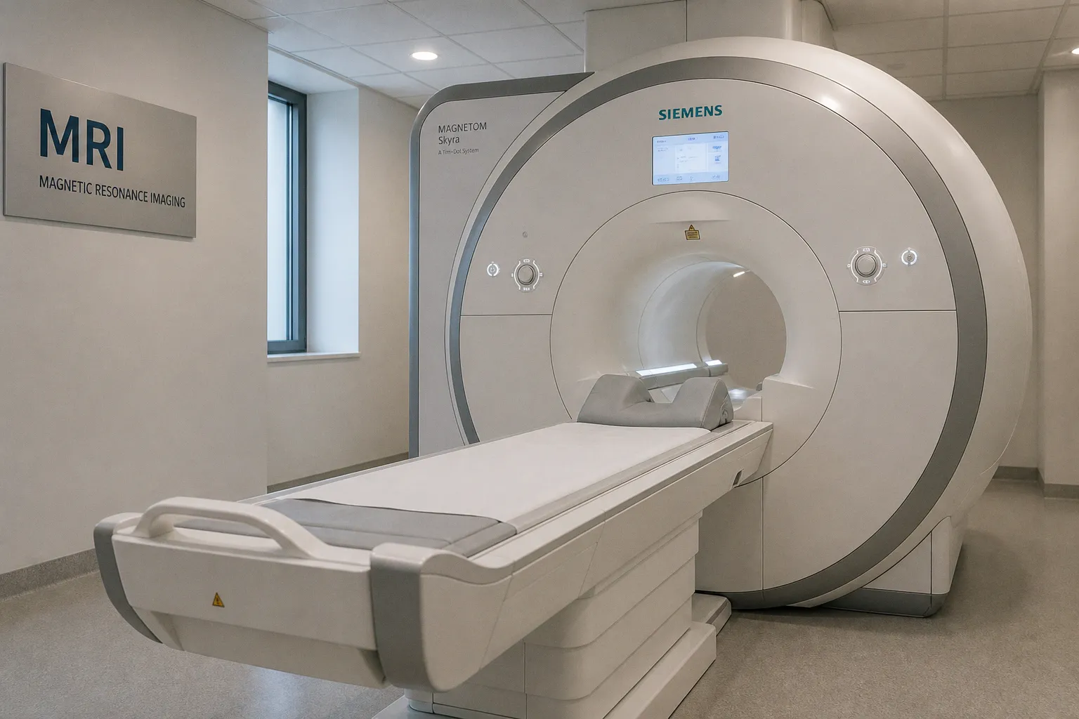
According to The Transmitter, a study published in Nature Neuroscience has found that structural MRI studies often fail to produce reproducible brain signatures for conditions including autism, depression, and bipolar disorder. Across independent datasets, measures such as cortical thickness and grey-matter volume showed little consistent agreement, raising a practical concern for anyone developing or evaluating neuroimaging biomarkers: a statistically visible regional difference is not necessarily a stable biological signal.
When the same contrast does not yield the same signature
Structural MRI has offered an appealing route into neuropsychiatry because it provides measurable features—cortical thickness, regional volume, and grey-matter distribution—that can be compared across groups. Yet the history of these studies has been marked by contradiction: some reports have described greater cortical thickness in selected regions among autistic participants, while others have reported the opposite.
The new analysis addressed whether these discrepancies might be explained primarily by differences in study design or image-processing methods. Researchers analyzed scans from thousands of people with neuropsychiatric conditions, including depression, schizophrenia, schizoaffective disorder, autism, and bipolar disorder, alongside scans from people with Alzheimer’s disease. They applied the same analysis pipeline across the datasets and compared whether structural changes were reproduced between independent sites.
The distinction was important. Alzheimer’s disease showed a robust and reproducible structural brain signature, whereas the neuropsychiatric conditions initially showed little agreement across studies. Adjusting for demographic and technical factors—including age, sex, and scanner differences—did not account for the inconsistency.
That result does not mean that structural MRI contains no information about neuropsychiatric illness. It does mean that traditional regional measures may not behave as reliable, general-purpose biomarkers for these diagnoses. Cortical thickness, in particular, carries an intrinsic amount of measurement error even under ideal conditions, according to commentary cited by The Transmitter.
The problem may begin before the scan reaches the algorithm
For neuroimaging software teams, the findings place the emphasis on reproducibility rather than novelty. A pipeline can be technically consistent and still produce an unstable disease signature if the underlying clinical categories contain broad and overlapping biological presentations.
The study’s authors point to a possible difference in diagnostic structure. Schizophrenia, for example, requires a consistent symptom pattern over six months, while autism, bipolar disorder, and depression encompass broader ranges of clinical presentation. If people grouped under one diagnostic label do not share a discrete structural pattern, averaging their scans may weaken or obscure any signal that exists at the individual level.
Consider the implications for model development. A regional effect that appears in one cohort should not be treated as a dependable biomarker until it survives independent-site testing, consistent processing, and assessment of measurement error. Scanner harmonization and demographic adjustment remain important, but this analysis suggests that they cannot, by themselves, resolve every source of variation.
This shift allows us to separate two questions that are often blended together: whether MRI can detect meaningful variation in the brain, and whether one regional measurement can reliably classify a broad psychiatric diagnosis. The first may still be productive; the second is where the evidence described here becomes notably more cautious.
What to watch next
The study supports a more restrained trajectory for structural MRI biomarkers in neuropsychiatry. Rather than expecting repeated group comparisons to converge automatically on a single cortical map, researchers may need approaches that better reflect clinical heterogeneity and individual variation—although the evidence provided here does not establish which alternative method will succeed.
For radiologists, neuroscientists, and developers, the immediate lesson is methodological: treat non-replication as information about the biomarker, not merely as noise to be engineered away. Claims built on cortical thickness or grey-matter volume should be read alongside their independent validation, diagnostic definitions, site differences, and known measurement limitations.
The broader clinical reality is quieter than the language of a “brain signature” can imply. MRI remains a powerful way to observe brain structure, but for many neuropsychiatric conditions, the path from scan contrast to diagnosis may not be a single regional measurement. A reliable trajectory will likely depend on acknowledging that complexity before promising a map.