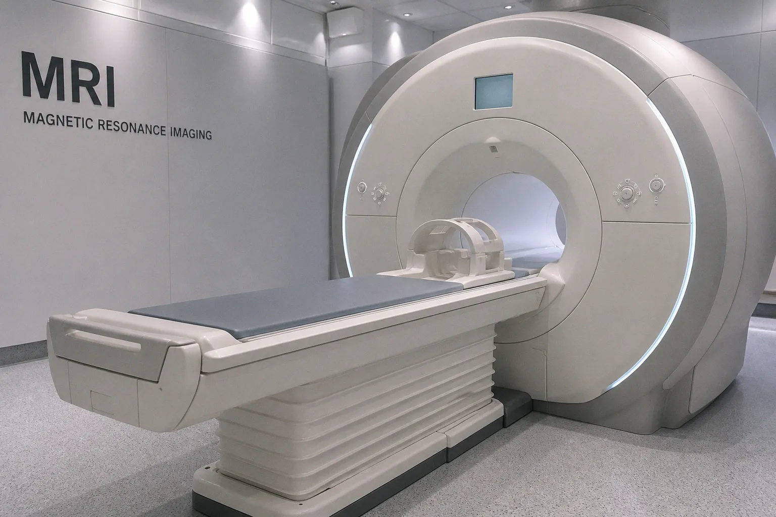
Drawing on MRI scans from 130 patients across multiple centers, the investigators report that an overwhelming 84.7% showed structural brain abnormalities, ranging from midbrain-hindbrain malformations to anomalies of the corpus callosum. For a field accustomed to fragmented case reports, the breadth of this dataset, and the biological pathway it traces, is what gives the work its weight.
What the scans actually show
The most arresting finding is not simply the prevalence figure but the pattern beneath it. Midbrain-hindbrain malformations and callosal anomalies together suggest that the dysregulation begins early, during the very scaffolding stages of neural development. Consider the implications: the imaging findings begin to function less as incidental observations and more as a developmental signature, one that maps onto the RAS/MAPK pathway's known role in shaping neuronal migration and midline patterning.
This shift allows us to think about Noonan syndrome not as a constellation of cardiac and facial features with occasional neurological involvement, but as a condition in which the brain itself is routinely part of the clinical picture. The multicenter design, pulling from heterogeneous scanners and protocols, also speaks to the reproducibility of the signal. Even with real-world variability in field strength and sequence selection, the abnormality rate holds.
From genotype to imaging phenotype
For the translational reader, the most useful framing is the bridge between genotype and phenotype that the study enables. The RAS/MAPK pathway, long implicated in the syndrome's craniofacial and cardiac manifestations, now has a corresponding structural footprint visible on standard MRI. That matters because it converts a molecular suspicion into something a radiologist can identify, measure, and follow along a longitudinal trajectory.
In practical terms, this strengthens the case for routine brain MRI in newly diagnosed patients, not as a screening reflex but as a way to establish a baseline against which subtle degradation can later be tracked. The protocol question, including which sequences to favor, which contrast choices to standardize, and how to harmonize reads across centers, is precisely the conversation that imaging engineers and clinical software teams should now be pulling in.
What to watch next
A few threads are worth following as the literature matures. The study's authors emphasize the clinical value of MRI without yet specifying how abnormal findings should alter management; that translational step, from scan to care pathway, remains open. The broader context reinforces the lesson. A real-world analysis of donanemab therapy found amyloid-related imaging abnormalities in 21% of treated patients, and recent diffusion MRI work in older adults has begun to characterize superficial white matter integrity as a buffer against the cognitive consequences of gray matter loss. Read together, these findings suggest that the scan is no longer an endpoint but the beginning of a trajectory the neuroimaging community is increasingly being asked to steward, with all the methodological care that stewardship implies.