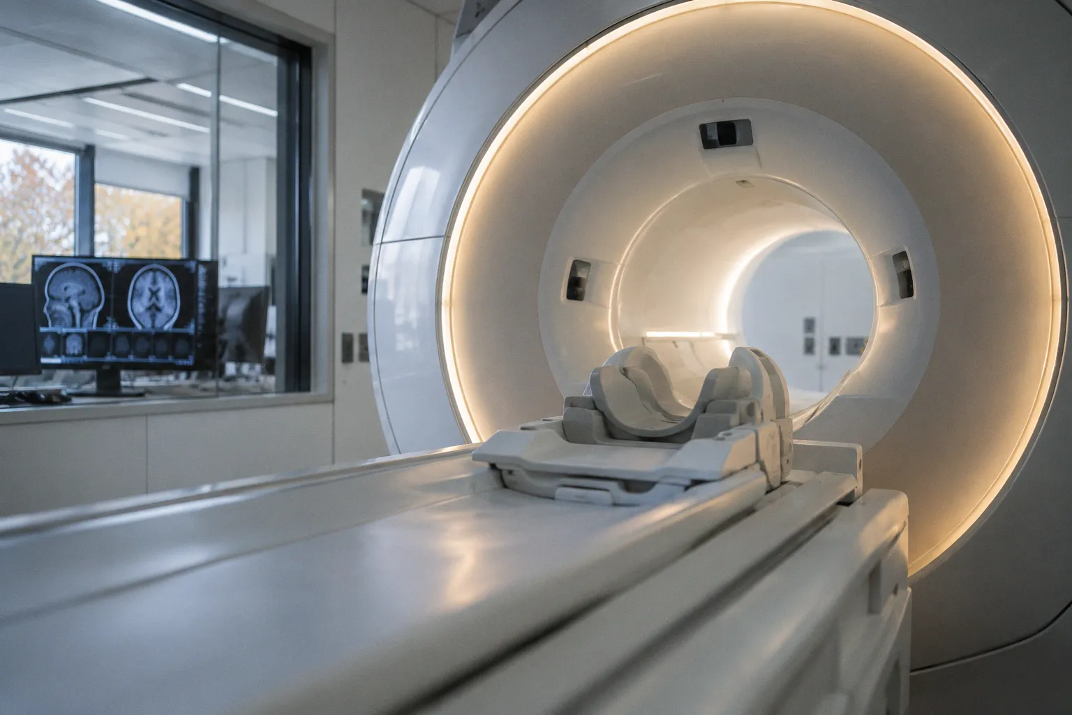
The work directly confronts the heavy-metal retention concern that constrains repeated imaging in neuro-oncology follow-up protocols. For algorithmic reviewers, the proposition is binary: does the synthesis preserve the diagnostic contrast gradient, or does it merely approximate it?
Where the architecture stops being described
The public framing reduces the model to a generative substitute operating in lieu of the contrast agent. The clinical claim — diagnostic quality maintained for glioma assessment — is the only substantive endpoint visible in current reporting. No SNR figures, no k-space trajectory specifications, no inference latency benchmarks, no multi-vendor generalization data appear in the available material. That omission is the binding constraint. The pipeline cannot be audited against standard acquisition physics until those numbers surface in a peer-reviewed venue with full reconstruction details. A generative network trained on contrast-positive input data inherits the contrast agent's noise floor along with its signal, and the published material does not yet specify whether the synthesis amplifies, suppresses, or simply reproduces that floor in the output domain.
What separates a deployment from a demonstration
Three checkpoints determine whether the system tolerates clinical workflow or remains a research curiosity. Lesion conspicuity must hold against histologically confirmed glioma margins, not against radiologist reader-study consensus alone, since the latter is known to tolerate visually convincing false negatives. Inter-scanner robustness must be demonstrated across at least two vendors and two field strengths — 1.5T and 3T — because the synthesis inherits every pulse-sequence imperfection from its training distribution. Failure-mode transparency matters most: when the model degrades on an unfamiliar morphology, the output must carry a calibrated uncertainty signal rather than a clean-looking false negative. Until those three conditions are satisfied, the synthesis yields a research artifact, not a replacement agent.
Reader-side verification checklist
For practitioners evaluating the work, the immediate question is provenance. Confirm the publication venue — a preprint tolerates different evidentiary standards than a peer-reviewed radiology journal. Confirm the training corpus composition: patient demographics, scanner vendors represented, glioma subtypes covered, and whether pediatric or recurrent cases were included in the cohort. Confirm whether gadolinium is removed from the acquisition protocol entirely or retained at reduced dose for ground-truth comparison purposes. Each of these details constrains the applicability envelope of the pipeline before any procurement or deployment decision is made.