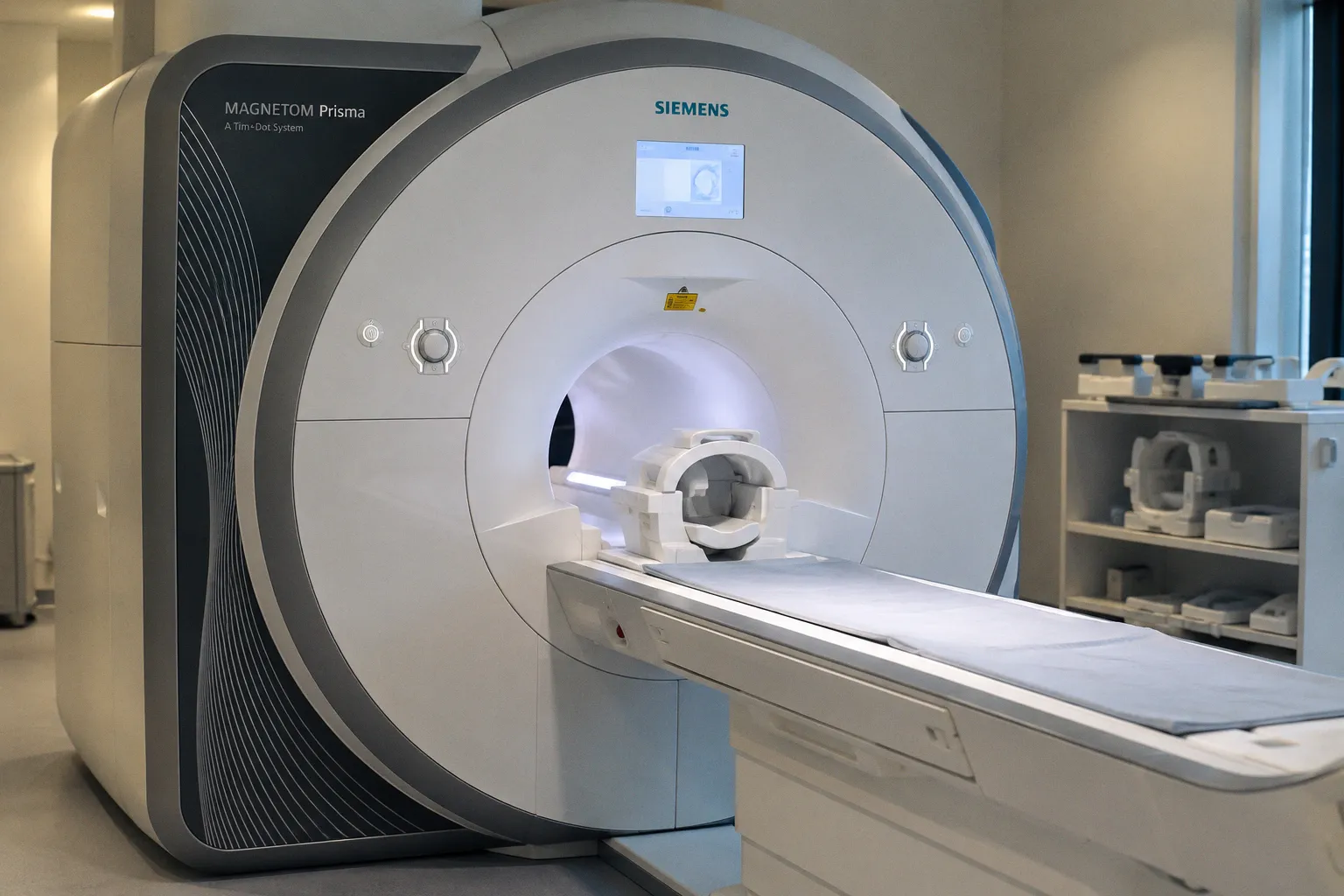
A new validation study posted to medRxiv asks a question that has lingered quietly at the intersection of radiology and computational neuroscience: when deep learning reconstruction compresses a T2-weighted brain MRI into a fraction of its original acquisition time, what happens to the volumetric maps that downstream segmentation tools produce from those images?
The validation question behind fast scans
The investigation focused on accelerated T2-weighted sequences processed through AI-based reconstruction algorithms, then fed into automated brain tissue segmentation pipelines. According to the medRxiv posting, the findings highlight how AI-reconstructed sequences influence structural neuroimaging volumetric precision, a subtlety that carries real consequences for longitudinal studies tracking subtle grey matter atrophy or white matter changes over months and years.
Consider the implications for a memory clinic following a patient through a five-year trajectory. If the reconstruction algorithm shifts the boundary between cortex and white matter even slightly from one scan to the next, the apparent rate of cortical thinning may begin to reflect the software rather than the biology. This is precisely the kind of measurement drift that erodes confidence in biomarker-based monitoring, and it is why careful validation work like this matters far beyond the technical audience who will read the preprint.
Speed gains and what they ask of us
The medRxiv study arrives alongside a related advance from Huashan Hospital, where radiology researchers have demonstrated the prospective feasibility of using deep learning reconstruction to deliver diagnostic-quality multi-contrast brain MRI in under 100 seconds. The two findings are complementary in a way that reframes the current moment in neuroimaging: acquisition is becoming dramatically faster, but every shortcut through the reconstruction network adds another layer at which volumetric measurements can quietly drift.
For translational researchers building segmentation pipelines, the practical takeaway is patience. Before swapping a DL-reconstructed sequence for an established conventional one, a clinic should run a small parallel comparison on representative cases, examine whether the resulting volumetrics remain within the tolerance their downstream analyses assume, and document the reconstruction kernel alongside every dataset. The temptation to celebrate the raw speed improvement is understandable, but in longitudinal and multi-site research the more durable metric is reproducibility of the biological signal across reconstruction versions.
Where the field is heading next
Meanwhile, the discipline is expanding outward in a different direction. A study from the University of Cape Town Neuroscience Institute reported the first use of point-of-care ultra-low-field MRI to examine the impact of antenatal maternal anaemia on infant neuroanatomy, speaking to the clinical utility of ultra-low-field neuroimaging for pediatric brain development assessments in resource-limited settings. On a separate axis entirely, work in Biological Psychiatry: Cognitive Neuroscience and Neuroimaging identified differential fMRI reward circuitry activation in resilient World Trade Center trauma survivors, with increased responses in the ventromedial prefrontal cortex and nucleus accumbens flagged as potential neural markers of psychological resilience.
What these threads share is a renewed attention to the assumptions baked into every reconstruction and analysis pipeline, and a recognition that the path from scanner output to clinical insight runs through software that deserves its own methodical validation.