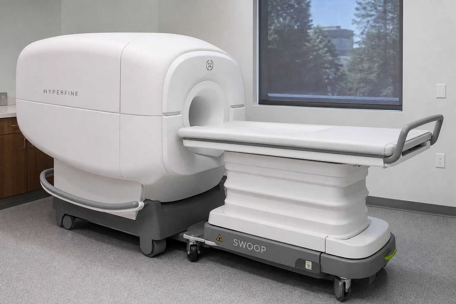
new study reported by AuntMinnie finds that Hyperfine's Swoop portable brain MRI system achieved 92% concordance with conventional high-field MRI when evaluating brain pathology — a result that, if it holds in broader clinical use, begins to reframe what imaging scientists and bedside clinicians should expect from low-field systems.
Reading 92% concordance with clinical care in mind
The headline figure invites careful interpretation rather than celebration. Portable scanners have long been constrained by signal-to-noise trade-offs that distort the subtle contrast differences on which radiological diagnosis depends, particularly in the cortical ribbon and deep gray matter structures where T1 and T2 relaxation properties shift the most. Consider the implications: when a low-field platform approaches the diagnostic agreement of a 1.5T or 3T scanner across a general pathology panel, it suggests that the acquisition and reconstruction pipelines have matured in ways that earlier iterations simply could not match. Still, concordance is not equivalence, and the unanswered question is how that remaining 8% distributes across lesion type, anatomical location, and patient population — the kind of nuance that determines whether a tool earns a permanent place in the imaging pathway or remains an adjunct for triage.
What portability changes for the patient trajectory
This shift allows us to imagine MRI as a longitudinal companion rather than a scheduled appointment routed through the radiology suite. For patients who cannot be safely transported — those on ventilators, with hemodynamic instability, in the early post-operative window, or in rural and emergency settings where a high-field magnet is hours away — the difference between a clinically usable scan and no scan at all is often the difference between a confirmed diagnosis and an educated inference. The Swoop's footprint quietly invites a workflow in which imaging follows the patient, rather than the patient being moved to the magnet, and that simple inversion carries real consequences for how quickly subtle degradation can be caught and acted upon.
Where the field stands this week
The encouraging trajectory sits alongside other recent developments worth keeping on the reading list. Bioengineer.org reports a deep learning reconstruction framework enabling free-breathing pediatric cardiac MRI that reduces scan time and the need for anesthesia; the University of Oxford has published work identifying two prefrontal-subcortical circuits that shape how we attribute outcomes to our own actions rather than to external circumstances; and MDPI Cancers describes how functional MRI during a silent verb generation task reveals activation changes in patients after radiotherapy. Taken together, the through-line is methodological rather than spectacular — software and reconstruction are doing the heavy lifting that used to demand longer scans, more sedation, or higher field strengths. For those of us watching the neuroimaging space, the practical question is no longer whether low-field portable imaging is technically feasible, but whether the concordance data generalize beyond the cohorts in which they were generated, and whether the imaging protocols we build around these tools preserve the diagnostic subtlety we are trained not to lose.