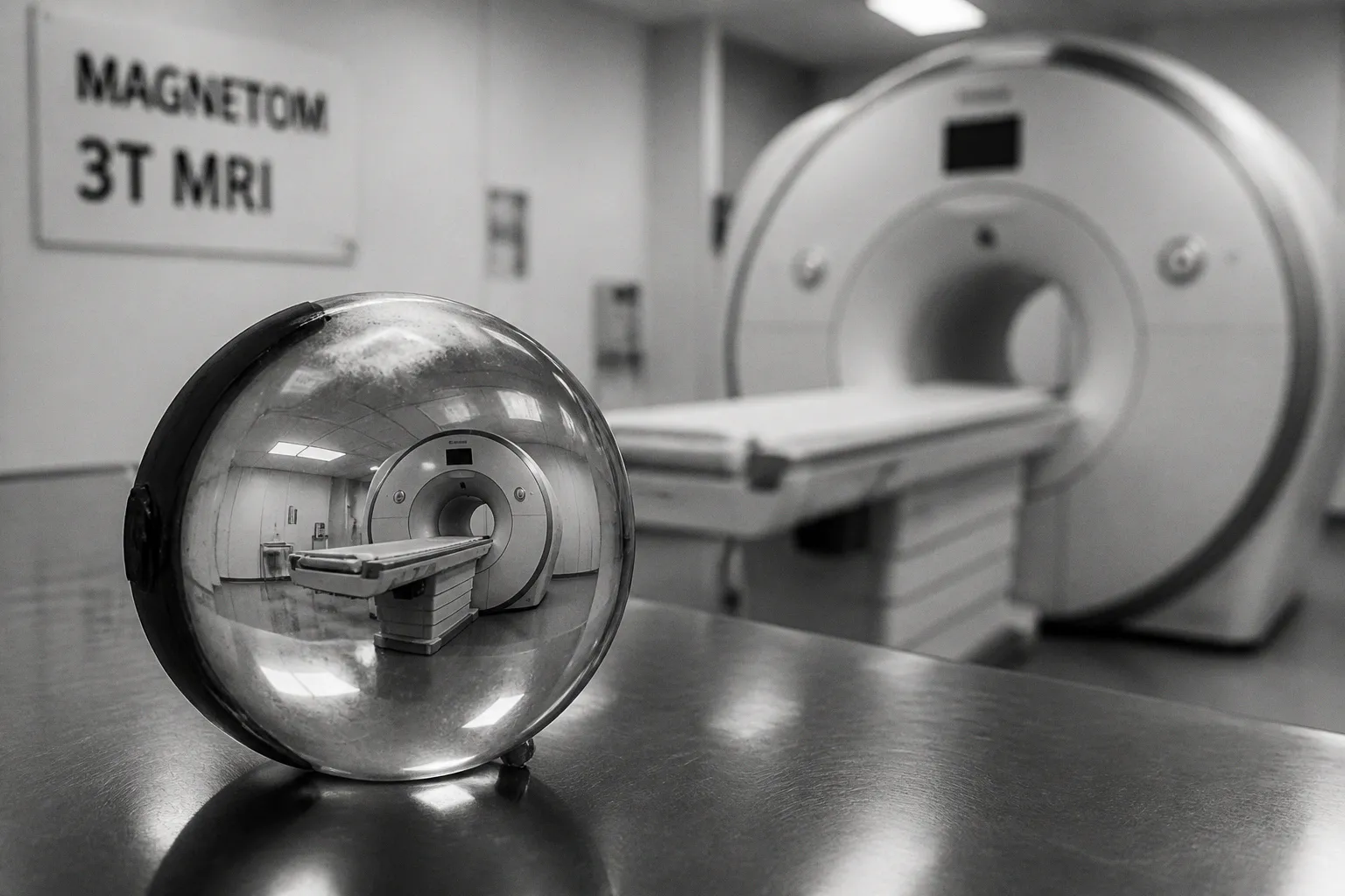
The footnote in fine print is the part nobody sees until the FDA sends a letter: deep learning reconstruction can hallucinate. Not in the LLM sense of fabricating sentences — in the more visceral sense of inventing structures that look like anatomy, or quietly erasing the small ones that actually matter.
The phrase "hallucination" in medical imaging has a more clinical ring than it does in generative text. Researchers use it to describe plausible-looking false structures produced by a reconstruction network, or — more dangerously — the silent deletion of subtle pathology that the model's training distribution did not sufficiently encode. Either failure mode lands on the same reading list: missed findings, false alarms, and an FDA that is finally paying attention.
What an AI hallucination actually looks like in the reading room
It does not announce itself. That is the operational problem with current generative reconstruction models — the bad outputs are designed to look identical to the good ones. The network has spent its training budget learning to produce images that a radiologist would find plausible, and once it has cracked that aesthetic, distinguishing a confident invention from a faithful reconstruction becomes a statistical, not perceptual, problem.
Two failure modes dominate the literature.
- Phantom structures. The network fills under-sampled regions with content consistent with its priors — a vessel where there isn't one, a smooth cortical ribbon where the original was noisy but diagnostically adequate, a pseudo-lesion that triggers an unnecessary follow-up.
- Silent erasure. The opposite failure. A faint hyperintensity that should have survived the denoising step gets averaged away because the model has learned that "normal brain" has no bright dots in that location. The radiologist sees a clean image. The patient has a microbleed.
The second is the worse failure clinically. A missed finding does not generate a second look. A fabricated one at least gets questioned.
A reconstruction that looks too clean is not the same thing as a reconstruction that is faithful. The first may have thrown away your pathology with the noise.
Why deep learning reconstruction is "highly unstable"
The phrase is not mine — it comes from a PNAS study out of the University of Cambridge and Simon Fraser University, where researchers demonstrated that aggressive undersampling combined with a learned reconstruction produces networks that are wildly sensitive to small input perturbations. A fraction of a millimetre of patient motion, a slight shift in the k-space trajectory, a minor coil calibration drift — any of these can shift the network from a faithful reconstruction to one with severe artifacts, or worse, to one that looks fine while quietly inventing detail.
The mechanism is not mysterious. Generative models interpolate. When you ask them to reconstruct from data that undersamples k-space by a factor of four or eight, they have no choice but to draw on what they learned during training. The question is whether the prior they learned is anatomically accurate enough to substitute for the missing measurements. For common anatomy, often yes. For the rare case, often no — and the model has no flag to tell you which mode it is in.
This is also why the instability compounds. If your input has even a small amount of variation that drifts the network out of its training distribution, you can land in regions of the latent space where the network produces confident but unreliable outputs. The Cambridge and Simon Fraser teams characterized these as "severe artifacts or significant errors." In practice, the failure looks like either a smeared patch that no radiologist would accept, or — the harder case — a perfectly presentable image where the network has simply chosen to draw something the data did not support.
The FDA finally has a metric for hallucination detection
The US Food and Drug Administration Center for Devices and Radiological Health, which has been watching the deep-learning imaging market with growing unease, has begun deploying regulatory science tools to detect exactly this class of failure. The most consequential of these is the scaled Fourier Ring Correlation, or sFRC — a metric adapted from localization microscopy and re-tooled for medical image restoration.
The intuition is straightforward. Any reconstruction has a spatial frequency spectrum. Beyond a certain cutoff, the input measurement carries essentially no signal — only noise. If a reconstruction claims high-frequency detail at spatial frequencies where the measurement has nothing to offer, that detail was invented. sFRC quantifies the boundary between measurement-supported content and prior-supported invention, and it does so at a resolution fine enough to be diagnostic.
This matters more than it sounds. Until recently, the quality metrics most vendors published were PSNR and SSIM — both of which compare the reconstruction to a ground truth averaged across the test set. They reward models that look like the average case. They do not penalize a model that is confident and wrong on the specific case in front of you. sFRC shifts the evaluation from "does this look like the population" to "does this image contain content the measurement actually supports."
| Metric | What it measures | What it misses |
|---|---|---|
| PSNR | Pixel-level similarity to ground truth | Confident per-pixel inventions that resemble the average case |
| SSIM | Structural similarity to ground truth | Local hallucinations consistent with global structure |
| sFRC | Frequency content supported by measurement vs. by prior | Does not identify which anatomical structures are invented |
| Faithfulness Score | Per-pixel alignment with measurement constraint | Requires a defined forward model and reference acquisition |
The takeaway for anyone evaluating vendor claims: ask which metric the FDA submission actually used. If the answer is PSNR or SSIM, the submission is measuring whether the images look nice, not whether they are faithful.
Mapping where the invention happens
A separate line of work from the Beckman Institute and the Computational Imaging Science Laboratory has produced a more elegant analytical tool: a decomposition of any reconstructed image into a measurement component and a null component.
The measurement component is the part of the image that the input data actually constrains. The null component is the part that lives in the null space of the forward operator — content the network is free to invent because the measurement does not pin it down. In a well-behaved reconstruction, the null component is small and the measurement component dominates. In a hallucinating reconstruction, you see the opposite: large, structured content in the null component, where the network has decided what should be there based on its training data.
The practical consequence is a hallucination map. Rendered as an overlay in the viewer, it shows the radiologist which regions of the reconstruction are data-supported and which are AI-supported. A lesion sitting in a high-null region is suspect by construction. The radiologist can either request a fully-sampled acquisition, or read the region with explicit awareness that the model is filling in blanks.
If the model is allowed to invent, it will. The only question is whether you can see where.
What actually mitigates this — and what doesn't
Three mitigation strategies have moved from theory into engineering practice, and none of them is a silver bullet. The honest summary is that they each buy something and cost something, and the right mix depends on which failure mode worries you most.
1. Physics-informed architectures. The network is given the MRI forward model — the Fourier transform, the coil sensitivity maps, the k-space trajectory — as a hard constraint. Rather than learning the physics implicitly from training data, it is forced to produce reconstructions that are consistent with the measurement operator by construction. This is the most architecturally invasive approach: it slows training, complicates deployment across vendor platforms, and forces closer collaboration between MR physicists and software engineers than the typical vendor is structured for. The upside is robustness — a physics-informed network cannot produce reconstructions that violate the measurement, which is the structural condition for most hallucinations.
2. Per-pixel uncertainty quantification. The network produces not just an image but a confidence map alongside it. Approaches range from Bayesian neural networks with explicit posterior distributions to ensemble disagreement, where the reconstruction is run through multiple stochastic forward passes with dropout masking and the variance between them is surfaced as a per-pixel uncertainty estimate. The confidence map is overlaid in the viewer, and the radiologist learns to treat low-confidence regions as needing either a closer look or a re-acquisition. The cost is reading time and cognitive load, both of which are already in deficit. The benefit is that the model becomes honest about its uncertainty rather than presenting everything with equal confidence.
3. Adversarial robustness training. During training, inputs are deliberately perturbed in ways designed to push the network out of its training distribution. The goal is to teach the model to degrade gracefully — produce a noisy, lower-confidence image rather than a hallucinated, high-confidence one — when it encounters out-of-distribution input. The empirical evidence suggests this improves worst-case behavior at the cost of average-case performance: the model looks slightly worse on the easy cases in exchange for failing less catastrophically on the hard ones.
The honest read is that none of these approaches is sufficient on its own. Physics-informed architectures prevent the grossest violations of measurement consistency but do not eliminate subtle prior-driven smoothing. Uncertainty quantification tells you where to be suspicious but does not, by itself, prevent the suspicious outputs. Adversarial training improves robustness to input shifts but cannot anticipate every shift a clinical scanner will encounter. The mitigation that actually works in production is the combination, layered — and even then, with a human radiologist in the loop who knows the failure modes.
The reading-room reality
Most deep learning reconstruction models currently deployed in clinical scanners were approved on the basis of equivalence studies — the vendor demonstrated that the AI-reconstructed images, when read by a panel of radiologists, produced diagnostic decisions concordant with the fully-sampled reference standard at an acceptable rate. That is a meaningful bar, but it is also a specific bar: it measures concordance on a curated test set, in a population selected to be representative of typical clinical practice, against a reference that itself is imperfect.
It does not measure what happens when the network encounters the long tail — the patient with unusual anatomy, the incidental finding that the training distribution did not include, the subtle lesion that lives at the edge of detection. For those cases, the AI-reconstructed image is being shipped into the reading room with the same confidence as the typical case, and the radiologist is being asked to distinguish between a faithful reconstruction and a hallucinated one with no flag, no confidence map, and no indication of which pixels are measurement-supported.
This is the operational reality behind the clinical trial warning. The technology is not banned. The FDA has not issued a halt. What regulators and the consensus groups are saying — increasingly, explicitly — is that the evaluation framework needs to catch up to the failure modes. Metrics like sFRC, frameworks like the null-space decomposition, and architectures like physics-informed networks are the tools being built to do that catching up. Until they are standard, the prudent posture for any radiology department is to know exactly which reconstruction model is on the scanner, what metric it was evaluated against, where its training distribution ends, and what the reading protocol looks like when the model encounters input it has not seen before.
A faster scan is not, by itself, a better scan. And a cleaner image is not the same thing as a faithful one. The reading room has always known this. The vendor deck is just catching up.