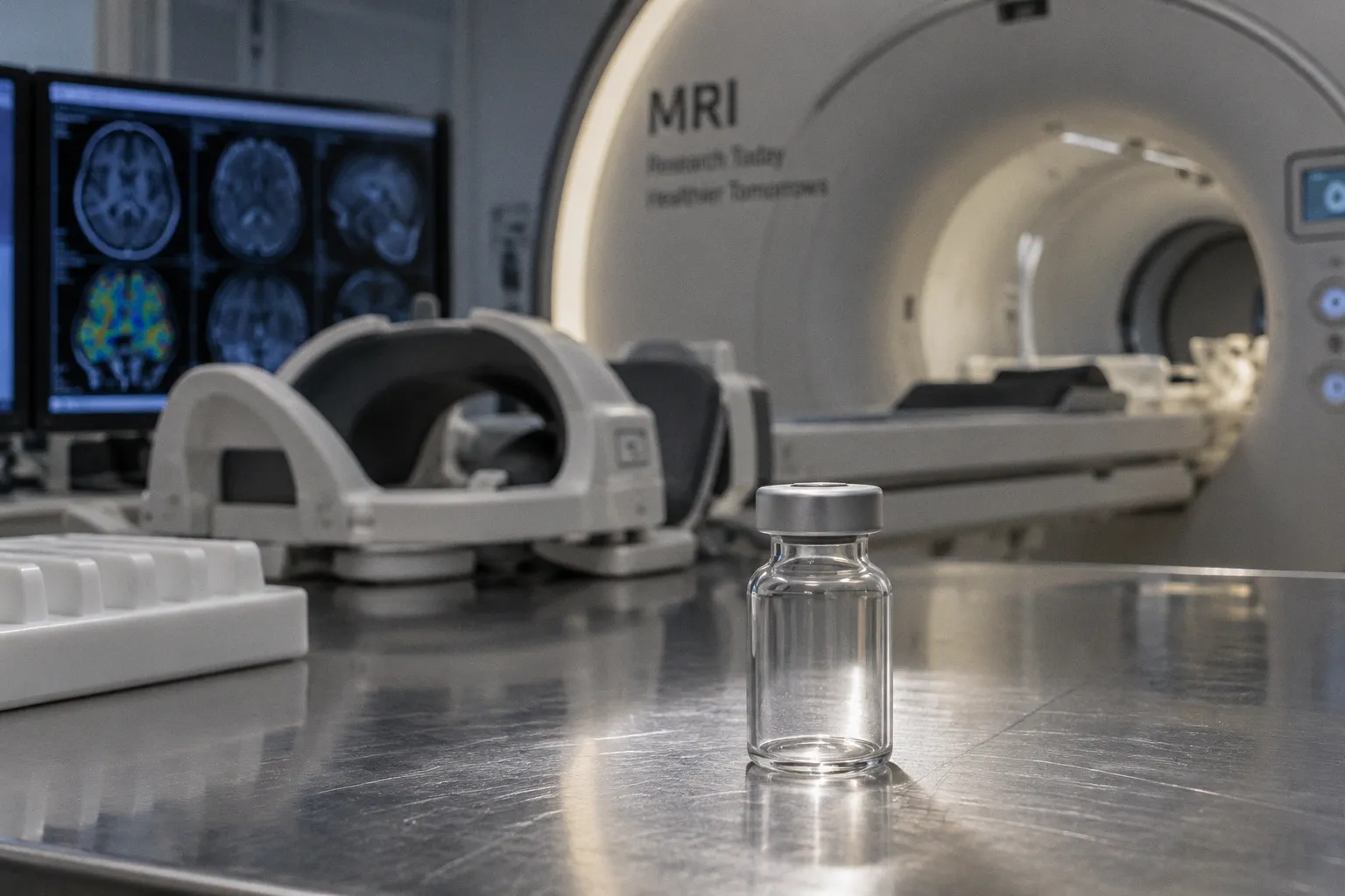
Cerebral blood flow, blood volume, vascular permeability, and contrast-agent kinetics are all clinically meaningful phenomena. The problem is that each is observed through a chain of acquisition choices, timing assumptions, reconstruction methods, and software implementations that can alter the final number before it reaches the trial database.
This is why a multicenter study may produce technically valid scans yet still struggle to demonstrate a reliable treatment effect. If one site measures relative cerebral blood volume from a carefully corrected dynamic susceptibility contrast examination and another uses a different preload strategy, distortion correction, arterial input function, or leakage correction, the resulting values may share a label without sharing the same measurement behavior. In clinical research, that distinction is not academic. It determines whether a longitudinal trajectory reflects subtle biological change or merely a shift in imaging practice.
Perfusion weighted imaging therefore needs to be treated as a measurement system rather than a single sequence. The system begins with the contrast between tissue and tracer, continues through acquisition and physiological modeling, and ends with a biomarker whose repeatability must be demonstrated before it is trusted as a clinical trial endpoint.
The first decision is biological: what does the study need to measure?
The phrase perfusion-weighted imaging covers several approaches that interrogate related but non-identical aspects of microvascular physiology. Dynamic susceptibility contrast MRI estimates changes in transverse relaxation during the passage of a gadolinium-based contrast agent, while dynamic contrast-enhanced MRI follows signal changes on T1-weighted acquisitions to model contrast delivery and leakage. Arterial spin labeling uses magnetically labeled arterial blood water as an endogenous tracer and can quantify cerebral blood flow without an intravenous agent.
These modalities should not be treated as interchangeable versions of the same test. They answer different biological questions, and the choice between them should follow the intended endpoint.
| Modality | Primary physiological signal | Common quantitative outputs | Main clinical research value | Principal limitation |
|---|---|---|---|---|
| DSC MRI | T2* signal change during contrast passage | Relative cerebral blood volume, perfusion-related measures, time-based parameters | Rapid assessment of hemodynamic contrast, particularly in neuro-oncology and acute ischemia | Sensitive to susceptibility effects, leakage, motion, timing, and preprocessing choices |
| DCE MRI | T1 signal evolution during and after contrast administration | Ktrans and related permeability or tracer-kinetic parameters | Characterization of vascular permeability, angiogenesis, and response to anti-angiogenic treatment | Requires kinetic modeling and reliable temporal sampling; results depend strongly on model assumptions |
| ASL | Labeling of endogenous arterial water | Cerebral blood flow | Repeatable, noninvasive hemodynamic monitoring without exogenous contrast | More vulnerable to transit-time effects, lower signal-to-noise ratio, and differences in labeling and post-labeling timing |
In an oncology trial, for example, Ktrans may be relevant because it reflects vascular permeability and capillary leakiness, features that can change under anti-angiogenic therapy. That does not make Ktrans a direct measure of tumor cell death, nor does a reduction necessarily establish durable clinical benefit. It is a biomarker of a vascular process whose meaning depends on the treatment mechanism, the acquisition protocol, and the surrounding clinical evidence.
In acute cerebral ischemia, the relationship between perfusion and tissue injury is more immediate. Combining perfusion-weighted imaging with diffusion-weighted imaging can help identify tissue that is hypoperfused but not yet irreversibly damaged, often described in the context of the ischemic penumbra. This distinction can support rapid treatment decisions within the standard three-hour intravenous thrombolysis treatment window. Yet even here, the image is not self-interpreting: the apparent mismatch depends on timing, motion, thresholding, and the specific analysis method used to define abnormal tissue.
A perfusion metric becomes a biomarker only when its biological meaning and measurement behavior are understood together.
DSC, DCE, and ASL begin with different assumptions
DSC MRI: fast passage, fragile interpretation
Dynamic susceptibility contrast MRI commonly uses a single-shot echo planar imaging acquisition with T2*-weighted contrast during the passage of a gadolinium-based agent. A standard dose often used for GRE-EPI DSC MRI is 0.1 mmol/kg. The sequence is efficient and well suited to capturing the first-pass bolus, which is why DSC remains central to many neuro-oncology and cerebrovascular protocols.
The familiar output is relative cerebral blood volume, or rCBV. In simplified terms, the passage of contrast causes a transient signal loss, and the area under the concentration-time curve is used as a proxy for blood volume relative to a reference region. The simplicity is deceptive. The observed signal is influenced by susceptibility, contrast recirculation, tissue geometry, bolus timing, venous contamination, and leakage of contrast into the extravascular extracellular space.
Contrast leakage is particularly important in tumors, where a disrupted blood-brain barrier can make the measured signal depart from the assumptions of an ideal intravascular tracer. Preload strategies, leakage correction, baseline definition, and the selection of normalizing tissue can all affect the resulting rCBV. A trial that specifies only the phrase DSC perfusion, without defining these elements, has not yet specified a reproducible biomarker.
In practice, the protocol should state the acquisition family, the timing of the dynamic series, contrast dose and injection conditions, temporal resolution, spatial coverage, and the intended correction and normalization approach. It should also make clear whether the endpoint is absolute or relative. Relative values may reduce some scanner-dependent effects, but they introduce their own dependence on reference tissue and segmentation.
DCE MRI: permeability is a model, not a raw signal
DCE MRI relies on repeated fast T1-weighted gradient-echo acquisitions during the administration of a paramagnetic contrast agent. The temporal resolution is typically in the range of three to six seconds per phase, allowing the study to follow contrast arrival, distribution, and washout over time.
The central quantities in DCE analysis are generated through tracer-kinetic models. Ktrans is commonly used to characterize the transfer of contrast from the plasma compartment into the extravascular extracellular space. Depending on tissue perfusion and permeability, Ktrans may reflect blood flow, endothelial permeability, or a combination of both. That ambiguity is not a flaw in the technique; it is a reminder that the parameter cannot be interpreted independently of the physiological regime and model used.
Two studies may both report Ktrans while using different arterial input functions, temporal windows, motion correction strategies, baseline T1 measurements, or kinetic models. Their numbers may not be directly comparable even when the same unit appears in the final report. This is one reason biomarker validation in oncology trials requires more than demonstrating that a metric changes after treatment. The study must establish whether the change is repeatable, biologically plausible, and sufficiently robust to distinguish treatment response from acquisition variability.
A useful DCE protocol therefore specifies the pharmacokinetic model in advance and identifies how arterial input will be obtained or estimated. It should also define how lesions are segmented, how necrotic or hemorrhagic regions are handled, and whether the endpoint is based on a mean, median, percentile, or spatially summarized distribution. Tumor heterogeneity is often biologically meaningful, but it can become analytical noise if segmentation and summary rules shift between time points.
ASL: endogenous labeling with a different set of vulnerabilities
Arterial spin labeling offers a distinct advantage: it measures cerebral blood flow using endogenous arterial water as the tracer, eliminating the need for an exogenous intravenous contrast agent. This makes ASL attractive for repeated examinations, for patients in whom contrast administration is undesirable, and for longitudinal hemodynamic monitoring where cumulative procedural burden matters.
The absence of contrast does not mean the absence of complexity. ASL is sensitive to labeling efficiency, post-labeling delay, arterial transit time, background suppression, motion, and the signal-to-noise characteristics of the sequence. A delayed arrival of labeled blood may be misread as reduced flow if the acquisition does not adequately account for transit time. Conversely, a protocol optimized for one patient population may behave differently in patients with vascular disease, collateral flow, or altered hemodynamics.
ASL should therefore be selected when its physiological strengths align with the trial question, not simply because it is noninvasive. It does not universally replace DSC or DCE in oncology studies. Its spatial and temporal characteristics, as well as its inability to provide the same permeability information as DCE, make it complementary rather than interchangeable.
Standardization starts before the first participant is scanned
Many multicenter imaging failures are established during protocol design, long before an endpoint is analyzed. A sequence name is not a protocol. The protocol is the complete set of conditions that determine how the signal is created, sampled, corrected, modeled, and summarized.
At minimum, a trial imaging charter should define:
- The physiological quantity of interest and the reason it is relevant to the therapeutic mechanism.
- The acquisition sequence, field strength, spatial resolution, temporal resolution, coverage, and duration.
- Contrast-agent identity, dose, injection rate, timing, and the relationship between injection and acquisition.
- Preprocessing procedures, including motion correction, distortion correction, denoising, and handling of signal outliers.
- Leakage correction, baseline correction, arterial input function strategy, and kinetic model where applicable.
- Lesion segmentation rules, reference tissue selection, and treatment of necrosis, hemorrhage, edema, or postoperative change.
- The primary summary statistic and the approach to missing, corrupted, or non-evaluable scans.
- Quality-control thresholds and the procedure for reviewing deviations before data are released for endpoint analysis.
This degree of detail can feel excessive when a trial is still focused on recruitment and clinical operations. Consider the implications, however, of discovering near the end of a longitudinal study that the early scans used one timing convention and later scans used another. A small protocol difference can become indistinguishable from a treatment-associated change, particularly when the expected effect is modest and the disease trajectory includes subtle degradation or fluctuating edema.
The imaging charter should also distinguish fixed elements from allowable adaptations. A site may need to use a vendor-specific implementation, but that flexibility should be documented, tested, and bounded. If every center is free to optimize the sequence independently, the trial has effectively created several related experiments rather than one harmonized measurement program.
QIBA profiles and the practical meaning of reproducibility
The Quantitative Imaging Biomarkers Alliance, established by the Radiological Society of North America, develops technical performance standards through QIBA profiles. These profiles are intended to improve measurement repeatability and reproducibility, particularly in multicenter clinical trials where the same biomarker must survive differences in equipment, operators, and local workflows.
Repeatability and reproducibility are related but distinct. Repeatability asks whether the same site, scanner, protocol, and processing pipeline can produce similar results under similar conditions. Reproducibility asks whether comparable results can be obtained across sites, scanners, operators, and implementations. A biomarker may perform well under repeatability testing while still failing when transferred to another vendor or institution.
For perfusion weighted imaging biomarkers in clinical trials, this distinction should shape the validation plan. A study may include test-retest examinations in a subset of participants, phantom or reference-object measurements where appropriate, and cross-site review of raw data and derived maps. The aim is not to eliminate all variation. Biological variation is often the signal of interest. The aim is to characterize technical variation well enough that a clinically meaningful change can be separated from measurement noise.
A robust validation program typically examines several layers:
1. Acquisition stability. Are the temporal resolution, coverage, contrast timing, and signal characteristics sufficiently consistent across repeated examinations?
2. Processing stability. Does the same input produce the same output when analyzed more than once or on different software installations?
3. Segmentation stability. Do different readers or automated methods identify comparable lesion volumes and regions of interest?
4. Cross-platform behavior. Does a nominally identical metric retain its meaning across scanner vendors and field strengths?
5. Longitudinal sensitivity. Is the metric responsive to biological change without being excessively responsive to small technical deviations?
This shift allows imaging scientists to discuss a biomarker in terms that are closer to clinical reality. A value is not useful merely because it is quantitative. It must also have a known error structure, a defensible biological interpretation, and a clear relationship to the decision the trial is intended to support.
DSC and DCE variability: where the numbers begin to drift
Timing and bolus delivery
Dynamic methods depend on time. In DSC, the relationship between the contrast bolus and the dynamic acquisition determines whether the first pass is captured cleanly. In DCE, temporal resolution influences the ability to distinguish rapid delivery from slower leakage and affects the stability of kinetic fitting. Injection timing, rate, line placement, and saline flush conditions can all influence the input function.
A protocol should not describe contrast administration as a procedural afterthought. It should define the timing relationship between injection and acquisition, identify acceptable deviations, and record deviations in a way that can be incorporated into quality control. When a bolus is delayed, dispersed, or partially compromised, the scan may remain visually interpretable while becoming unsuitable for a quantitative endpoint.
Leakage and susceptibility correction
DSC is especially vulnerable when the blood-brain barrier is disrupted. In an enhancing tumor, extravasated contrast can alter the signal beyond the intravascular susceptibility effect that the basic model assumes. Leakage correction can reduce this bias, but the correction itself depends on the implementation and on the assumptions used to estimate the contaminating signal.
DCE has the opposite interpretive direction: leakage is part of the target phenomenon, but its quantification depends on a model that separates delivery, permeability, and extracellular distribution. In both cases, the word correction should not imply that the underlying problem has disappeared. It means that a defined mathematical strategy has been applied, and that strategy must be kept consistent and validated.
Region-of-interest design
Perfusion metrics are often summarized over a lesion, but lesions are not uniform. A tumor may contain viable enhancing tissue, necrosis, hemorrhage, infiltrative margins, treatment-related change, and regions with markedly different vascular behavior. A whole-lesion mean may conceal the most responsive subregion, while a maximum value may be driven by noise or by a small area of atypical biology.
The analysis plan should make the segmentation logic explicit. If the trial uses enhancing tumor only, that boundary must be defined consistently across time. If edema or necrosis is excluded, the exclusion criteria should be operational rather than interpretive. If the endpoint is based on percentiles or histogram features, those choices should be justified in relation to the biological question and locked before outcome analysis.
The role of ASL in longitudinal monitoring
Longitudinal studies place a premium on tolerability and repeatability. ASL can be valuable in this setting because it quantifies cerebral blood flow without repeated administration of an intravenous contrast agent. This is particularly relevant when participants undergo multiple scans over months and the research question concerns a gradual trajectory rather than a single treatment milestone.
Yet the same longitudinal design exposes ASL to physiological confounding. Hydration, carbon dioxide levels, medication, blood pressure, alertness, and recent activity can influence cerebral blood flow. If these variables are not controlled or at least recorded, a change in CBF may be difficult to interpret. The issue is not that the measurement is invalid; rather, the biological system is responsive to more than the disease process under investigation.
Post-labeling delay deserves specific attention. If labeled blood has not reached the tissue at the time of readout, the measured signal can underestimate flow. A single delay may be adequate in one population and insufficient in another, especially where arterial transit is prolonged. Multi-delay approaches can provide additional information but may increase acquisition time and alter the operational burden of the protocol. The choice should be driven by the expected vascular physiology and the endpoint requirements.
ASL can also serve as a complementary measure alongside DSC or DCE. Concordant changes across methods may strengthen confidence in a hemodynamic interpretation, while discordant results can reveal that the modalities are measuring different components of the vascular response. That is not necessarily a problem to be averaged away. It may be the most informative result in the study.
The goal of harmonization is not to make every scanner identical; it is to make every difference visible, bounded, and interpretable.
Inter-vendor discrepancies do not end with acquisition
Even when acquisition protocols are carefully aligned, post-processing can introduce substantial divergence. Software may differ in how it converts signal-time curves into contrast concentration, identifies the baseline, handles negative or noisy values, estimates an arterial input function, performs leakage correction, or fits a kinetic model. Two pipelines can therefore produce different biomarker maps from the same raw series.
For multicenter research, the preferred strategy is usually to reduce unnecessary degrees of freedom. A centralized processing pipeline can improve consistency, provided that raw data are transferred with sufficient metadata and that the pipeline can accommodate vendor-specific image organization without silently changing the analysis. If local processing is necessary, the trial should establish version control, parameter locking, audit trails, and a mechanism for cross-site concordance testing.
A practical harmonization program may include:
- A common acquisition template translated into vendor-specific implementation instructions.
- Central review of pilot scans before the first participant enters the efficacy population.
- Automated checks for missing dynamics, incorrect coverage, motion burden, contrast timing, and signal dropout.
- Locked software versions and documented parameter files.
- Blind reprocessing of a shared data subset to compare site-level outputs.
- A predefined rule for labeling scans as usable, usable with qualification, or non-evaluable.
- Storage of intermediate maps and quality metrics rather than only the final biomarker value.
These steps matter because a final number without its provenance is difficult to defend. Clinical trial imaging endpoints need an evidentiary chain: the scan was acquired as specified, the data passed quality control, the processing followed a documented method, and the resulting metric retained acceptable behavior under repeated and cross-site testing.
From technical metric to trial endpoint
A perfusion metric should not become a primary endpoint simply because it responds to treatment earlier than a conventional clinical measure. Early change may be useful, but it can also reflect transient vascular normalization, altered permeability, edema reduction, or changes in contrast delivery that do not correspond to durable tumor control or functional recovery.
The endpoint framework should therefore connect four elements:
1. Mechanism: What vascular or hemodynamic process is expected to change under the intervention?
2. Measurement: Which PWI modality and parameter most directly capture that process?
3. Validation: What evidence shows that the parameter is repeatable, reproducible, and sensitive to relevant change?
4. Clinical interpretation: How will the imaging result be interpreted alongside symptoms, structural MRI, laboratory measures, progression criteria, or survival?
In oncology, for instance, a change in Ktrans may indicate altered permeability and angiogenic activity, but it should not be treated as a standalone proof of response. In neurovascular disease, perfusion and diffusion may clarify tissue status during an acute event, yet the imaging interpretation still belongs within a time-sensitive clinical pathway.
The same discipline applies to radiogenomics and molecular imaging. More complex models may reveal associations between imaging phenotype and genotype, but complexity does not remove the need for measurement control. A highly expressive algorithm trained on inconsistently acquired perfusion data can learn site characteristics as easily as it learns biology. Without harmonized inputs and external validation, the apparent sophistication of the model may conceal a fragile biomarker.
A more durable way to design PWI studies
The most reliable perfusion imaging programs are built around a modest but demanding principle: define the measurement before asking it to carry a clinical conclusion. That means selecting the modality according to the biological question, specifying the acquisition in operational detail, testing repeatability and reproducibility, and preserving enough provenance to understand every derived value.
For investigators planning a multicenter trial, the sequence of decisions is consequential:
1. Start with the vascular biology and the anticipated treatment effect, not with the availability of a familiar sequence.
2. Select DSC, DCE, ASL, or a complementary combination according to the physiological quantity required.
3. Lock acquisition, contrast, timing, segmentation, modeling, and processing choices before endpoint analysis.
4. Use QIBA-aligned principles and site qualification to establish measurement behavior across the network.
5. Treat quality control as part of the endpoint, not as an administrative filter applied after the study is complete.
6. Report limitations openly, particularly where vendor, model, or physiological differences remain unresolved.
Perfusion weighted imaging is already capable of supporting more precise clinical research, but precision does not arrive automatically with a color map or a decimal value. It is earned through protocol discipline, biological reasoning, and longitudinal attention to how a measurement behaves in real patients over time.
The central task is not to force DSC, DCE, and ASL into a single universal language. It is to understand what each modality is saying, control the conditions under which it speaks, and ensure that a change in the final metric reflects the patient’s biology more often than it reflects the machinery of measurement.