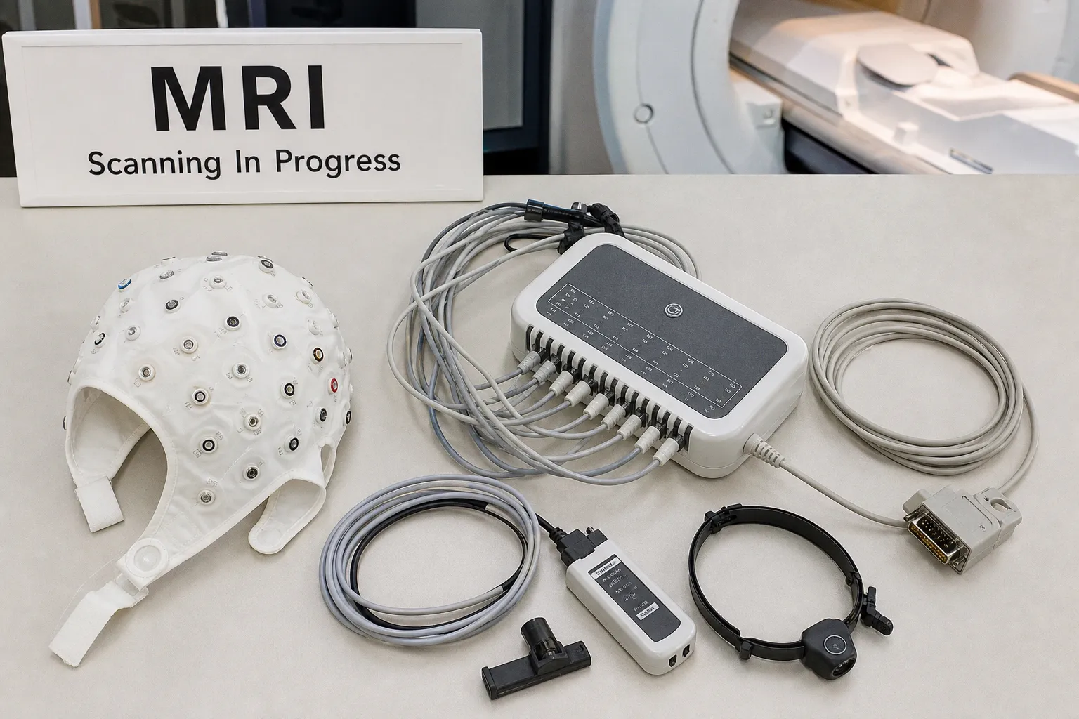
Researchers at Monash University have spent five years assembling the most extensive single-site dataset yet on how psilocybin reshapes functional brain connectivity — and, according to the report, how that reshaping depends on the environment in which the drug is administered. The clinical promise of psychedelic-assisted therapy has long outpaced the imaging evidence beneath it, which is precisely why a study of this scale and duration warrants careful attention from anyone working at the intersection of MRI methodology and translational neuroscience.
What the five-year timeline actually buys you
The duration matters as much as the headcount. Five years of acquisition at a single site implies a level of protocol standardization that multicenter psychedelic research rarely achieves — the same scanner, the same sequence upgrades, the same preprocessing lineage, year after year. When the signal of interest is as context-sensitive as functional connectivity, that kind of consistency is not cosmetic; it is the difference between a finding you can trust and one that drifts with every software update. Pairing that stability with AI-driven analyses capable of handling dynamic, time-resolved signals points to a methodological posture that treats psilocybin's effect on the connectome not as a static signature but as a trajectory worth following.
Context as a covariate, not a backdrop
The most intriguing thread, and the one most relevant to protocol designers, is that reorganization appears to track environmental context. For anyone building imaging sessions around psychedelic-assisted therapy, this reframes the scanner room itself as part of the experimental condition rather than a controlled neutral space. A standard fMRI suite is acoustically loud, spatially constrained, and stripped of the therapeutic framing that observational trials have associated with favorable outcomes. If connectivity shifts co-vary with setting — even modestly — then any imaging study of psilocybin must reckon with the mismatch between the scanner bore and the clinical room. It also opens a methodological question worth sitting with: should future protocols attempt to soften the scanner environment, or accept that contrast as a feature of the experimental design rather than a confound to be minimized?
What remains to be clarified
Several practical questions are not yet visible in the public reporting. How "environmental context" was operationalized — through deliberate manipulation, correlation with participant self-report, or inference from imaging features alone — will determine how replicable the result proves to be across labs. The precise role of the AI component, whether it functioned primarily as a classifier of connectivity states or as an analytic engine for extracting dynamic features, will shape how the work is received by the neuroimaging software community and which pipelines it ultimately feeds into. For now, the study signals something durable: a single research group has committed half a decade of imaging infrastructure to mapping how a psychoactive compound reshapes the human connectome under real conditions. That is the kind of longitudinal foundation the field has been waiting for — and exactly the dataset that will take years, not months, to fully interpret.