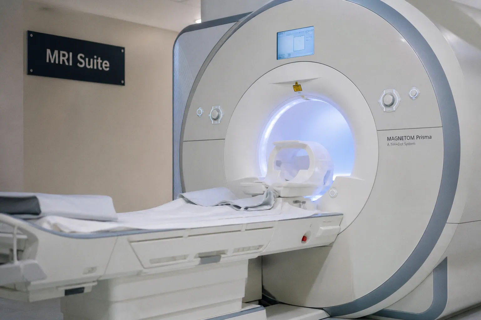
When the Rim of the Lesion Tells Two Stories
The study, as reported, places magnetic resonance spectroscopy (MRS) side by side with the more familiar post-contrast MRI, asking how each modality performs when a radiologist encounters that characteristic enhancing rim on a scan. For clinicians who routinely sit with these images, the question is not academic — the differential diagnosis behind a ring-enhancing lesion is wide, and the choice of sequence can quietly shape what comes next at the tumor board.
Why a Ring-Enhancing Lesion Defies Easy Answers
Consider the implications of a single contrast-enhancing rim on T1-weighted imaging. Beneath that bright border, the tissue can harbor necrotic tumor, an active inflammatory abscess, a resolving hematoma, or a demyelinating process whose own biology mimics neoplasm. Post-contrast MRI captures the breakdown of the blood-brain barrier — a vascular signature — but it does not directly interrogate the metabolic state of the tissue inside the ring. This is precisely the gap the Cureus analysis appears designed to address, framing MRS as a complementary probe rather than a competitor.
In practice, MRS contributes a different kind of evidence: ratios of metabolites such as choline, creatine, N-acetyl aspartate, and lactate, each shifting in patterns that trace the underlying cellular trajectory. When the contrast rim shows enhancement but the spectroscopy reads neoplastic metabolism, the clinician gains a second axis of confidence. When the two disagree, the case demands a longer conversation and, often, follow-up imaging along the longitudinal arc rather than an immediate procedural decision.
What the Field Is Watching Alongside It
The Cureus paper lands in a week when several adjacent threads are also moving through the neuroimaging literature. MDPI's Tomography has published a controlled pilot study using functional MRI, cortical thickness analysis, and diffusion tensor imaging in patients with persistent auditory verbal hallucinations, identifying structural and functional network differences centered on the frontotemporal and default mode networks. Separately, work highlighted by Tech Times points toward identifying the brain's language network from routine scans — a development with clear relevance for stroke patients who cannot tolerate lengthy task-based paradigms. And in basic neuroscience, Nature carries a study describing a reusable modular architecture that enables flexible cognitive operations in both mouse brain and artificial recurrent networks, a reminder that the algorithmic vocabulary shaping MRI analysis pipelines continues to evolve from biological inspiration.
Taken together, these pieces sketch a field that is no longer satisfied with a single sequence telling the whole story. For the ring-enhancing lesion specifically, the practical takeaway is unhurried but worth absorbing: post-contrast MRI remains the workhorse for detection, while MRS offers a metabolic reading that can either reinforce or complicate the vascular one. Clinicians building their protocols should consider where spectroscopy fits into their local workflow — not as a replacement for contrast imaging, but as a second voice in the diagnostic dialogue, one that speaks the language of cellular chemistry rather than vascular permeability.