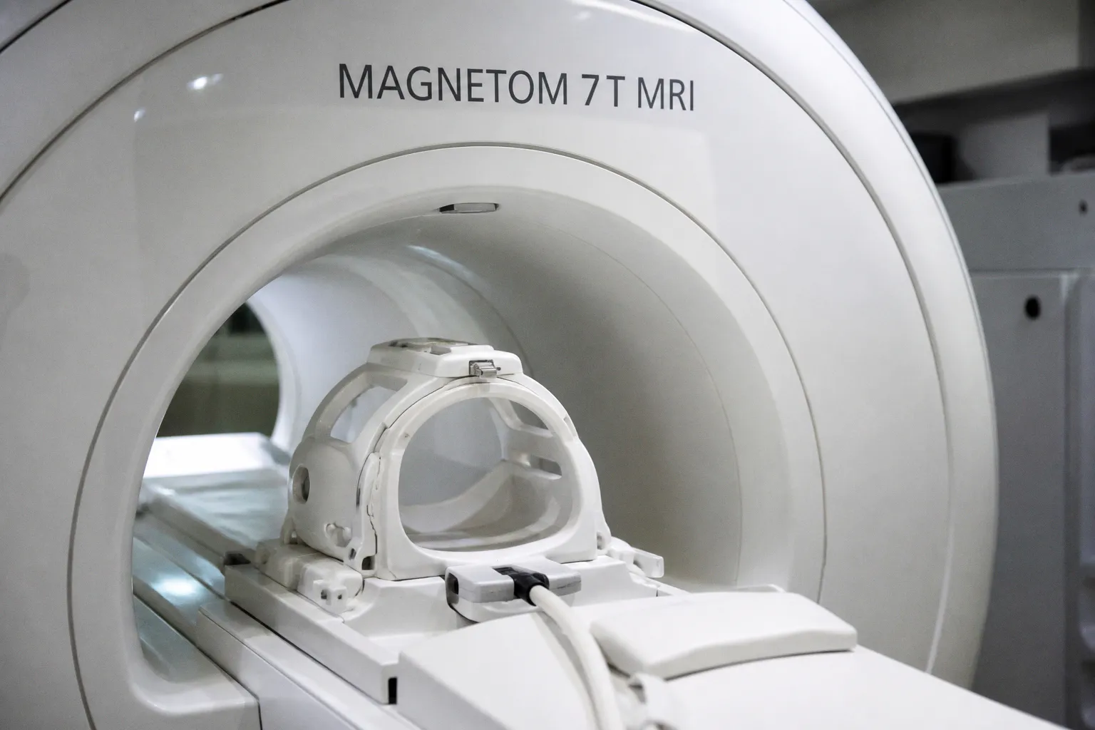
A study published in Nature Communications, led by researchers at National Taiwan University and the University of Illinois at Urbana-Champaign, has used 7-Tesla MRI to resolve submillimeter gray matter nuclei involved in high-stakes, value-based decision-making. By tuning their lottery decision task to the resolution limits of ultra-high-field imaging, the team identified the locus coeruleus and layered frontal cortex circuits as functional gates during choice formulation and outcome evaluation, offering a level of anatomical precision that conventional 3-Tesla protocols simply cannot reach.
Why 7T matters for the mesoscale
Consider the implications of moving from a standard 3-Tesla scanner, which resolves neural activity at roughly 3 mm, to a 7-Tesla system that can distinguish structures ranging from hundreds of microns to a full millimeter. This shift allows us to peer into the mesoscale architecture of the human brain, the intermediate territory between broad functional networks and single-cell recordings, where most of clinical neuroscience has historically worked with educated inference rather than direct observation. The researchers redesigned their stimuli specifically to engage these smaller hubs, a methodological choice that proved decisive. "By tuning our task stimuli to specifically target mesoscale structures at 7T, we could disentangle a series of small structural hubs in the brain that act as functional gates for different kinds of information during a decision," explains co-corresponding author Dr. Joshua Goh, an associate professor at NTU's Graduate Institute of Brain and Mind Sciences. "At 3T resolution and with less optimized tasks, these fine-grained functional distinctions would likely remain hidden."
A layered cortex under the scanner
The findings trace a coherent biological pathway from the brainstem upward. When salient high stakes entered the task, the locus coeruleus, a slender brainstem structure roughly 2 mm across, signaled the frontal cortex to allocate heightened neural resources rather than continuing at baseline operating levels. Within the 3 mm-thick frontal cortex itself, engagement shifted with remarkable specificity: deeper layers activated during the formulation of a choice, while superficial layers engaged when participants evaluated the outcome. This depth-resolved functional separation, captured non-invasively in humans, is the kind of result that was previously confined to invasive animal recordings or postmortem histology. It reframes the frontal cortex not as a single decision-making module but as a vertically organized sequence of processing stages, each contributing to a different phase of the choice trajectory.
What this opens for clinical practice
For clinicians and software developers working at the interface of imaging and patient care, the practical takeaway is twofold. First, the study underscores why 7-Tesla acquisition protocols and the analysis pipelines built around them deserve continued investment, particularly as neuroimaging software evolves to handle the motion-correction and distortion challenges that come with submillimeter acquisitions. Second, it sharpens the conversation about maladaptive decision-making in neuropsychiatric and neurodegenerative conditions, where a precise understanding of which nuclei are misfiring at which stage of the choice process could eventually guide targeted interventions, whether pharmacological, neuromodulatory, or cognitive. As with any single imaging biomarker, the temptation to overinterpret should be resisted; however, the methodological demonstration itself, that the human brain's decision hubs can be mapped at this resolution, is a meaningful step along the longitudinal arc of translational neuroscience.