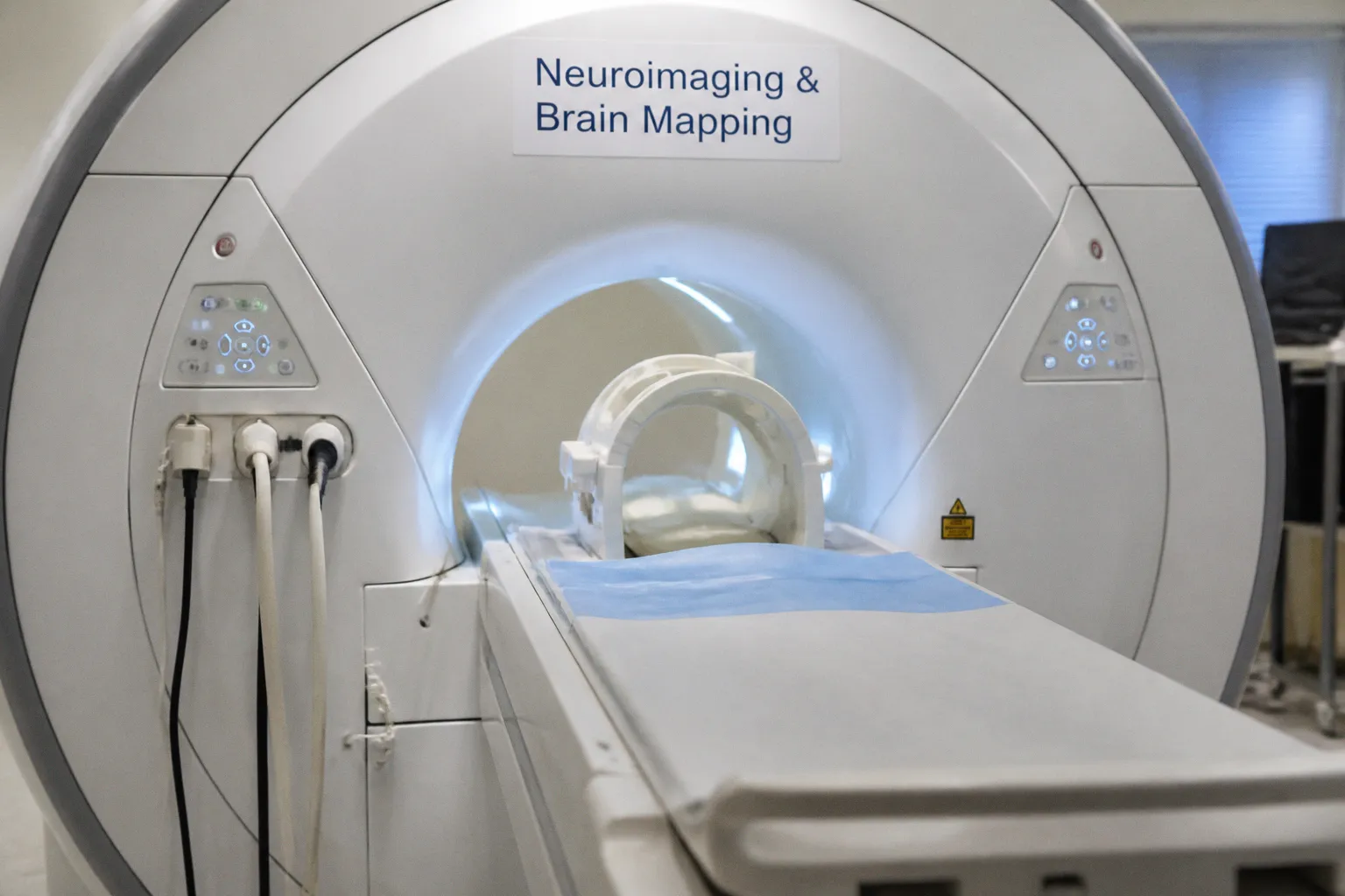
The lungs need protection from the inflammatory cascade that defines acute respiratory distress syndrome, and so does the brain — yet the very strategies we use to spare the lung can shift cerebral perfusion, raise intracranial pressure, or change how a follow-up MRI should even be read. It is precisely this tension that Bioengineer.org has turned its attention to in a recent piece on managing ARDS in the context of acute brain injury, and for anyone working at the seam between neuroimaging software and clinical practice, the discussion lands close to home.
Where lung strategy meets the scanner's readout
In the ICU, the conversation between the pulmonologist and the neurologist has always been a quiet one, conducted through tidal volumes and cerebral perfusion pressures rather than words. The framing offered by Bioengineer.org leans into this overlap, and it matters here because every ventilator adjustment leaves a downstream signature on the imaging studies we eventually run. Consider the implications: a patient who is hyperventilated to manage intracranial pressure will show altered cerebral blood flow patterns on subsequent perfusion imaging; a patient managed with higher positive end-expiratory pressure for oxygenation may present with subtle changes in venous drainage that complicate the interpretation of susceptibility-weighted sequences. For developers building neuroimaging pipelines, this means that the clinical metadata accompanying a scan — ventilator settings, sedation depth, the timing of neuromonitoring relative to image acquisition — is no longer a footnote but a critical variable that quietly shapes the signal we extract.
What remains unclear, and what to watch
The available material frames the question without yet resolving it, and that ambiguity is worth honoring rather than rushing past. We do not yet have a consolidated evidence base that tells the neuroimaging community which ARDS protocols consistently preserve the integrity of brain-imaging biomarkers, nor do we have standardized reporting of how ventilator parameters propagate into quantitative MRI outputs. This is where the translational neuroscience community has room to contribute: the same long, careful thinking that has refined diffusion-based measurements in chronic neurodegeneration now needs to be applied to the acute, mechanically ventilated brain, where the imaging substrate is being reshaped by clinical decisions made hours before the scanner is ever switched on.
A clinical lens for the software bench
For those of us who translate between the scanner console and the bedside, the practical reminder is straightforward. When an imaging study comes off the MRI in a patient recovering from acute brain injury and concurrent ARDS, the temptation is to read the image as a clean snapshot of the brain alone. But the trajectory of that scan reflects every breath the patient has been given. Building software that can flag — rather than silently absorb — these upstream clinical decisions is part of how we honor the full biology of the person on the table, and part of how we move neuroimaging from a window onto the brain toward a more honest mirror of the whole patient.