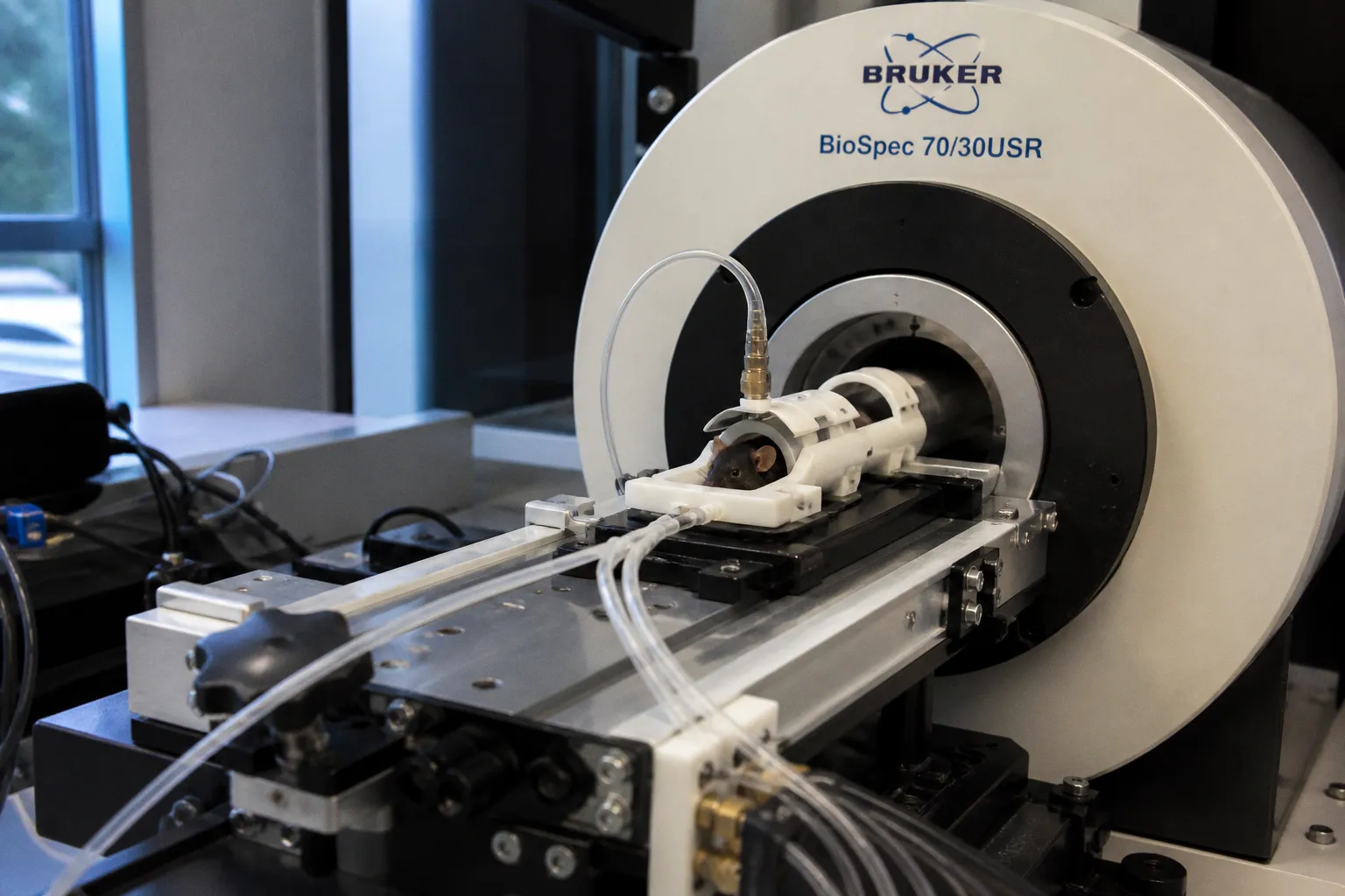
By showing that identical patterns of neural activity can produce radically different laminar blood flow responses across the cerebral cortex, the researchers make a case that the vasculature is not merely plumbing beneath the signal — it is part of the circuit. Consider the implications for every pipeline that treats the BOLD response as a faithful proxy for neuronal firing: the architecture of the vessels themselves shapes what we see, and that architecture is not uniform from layer to layer.
What the cortex does differently between pain and touch
In the mouse model the team examined, sensory inputs that arrive through distinct pathways — the sharp, alerting channel of pain and the softer, discriminative channel of touch — engaged overlapping neural ensembles yet recruited the surrounding microvasculature in visibly unequal ways. This shift allows us to step back from the assumption that the hemodynamic blanket reflects neural activity with the same fidelity across conditions; the local geometry of capillaries and penetrating arterioles appears to amplify, dampen, or redistribute the signal before it ever reaches the scanner. For anyone building preprocessing pipelines or layer-specific analysis tools, the practical message is that condition, stimulus type, and cortical depth all modulate the vascular contribution to the BOLD readout.
Why this matters for fMRI software and protocol design
The translational consequence runs deeper than a methodological footnote. If vascular architecture behaves as an active circuit component, then generic hemodynamic response functions — the fixed shapes many software packages still default to — may be biasing group comparisons, particularly in studies that compare patient populations where vessel health differs. Neurodegenerative cohorts, aging populations, and anyone with vascular risk factors sit precisely at the intersection where this caveat carries weight. A careful radiologist or neuroscientist scanning this literature should ask whether their acquisition protocol samples at a resolution that can resolve laminar differences, and whether their analysis stack conditions the hemodynamic model on stimulus type or vascular territory rather than applying a one-size-fits-all kernel.
What to watch next
The full mechanics — which cortical layers, which stimulus contrasts, and which downstream analytic adjustments the authors propose — are worth tracking as the paper moves through peer review and supplementary materials. For the software side of our readership, the open question is whether major toolkits will absorb condition-specific hemodynamic models or expose the vascular contribution as an explicit covariate. The biology has already given its answer; the engineering response is the part of the story worth watching closely.