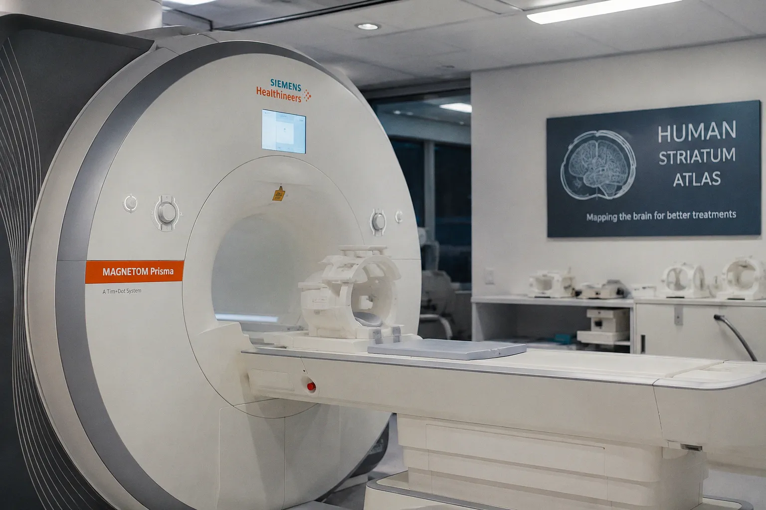
According to MIT News, researchers at the Massachusetts Institute of Technology have created a new cellular atlas of the striatum, identifying 31 neuronal subgroups through single-cell RNA sequencing and other techniques. The resource links distinct gene-expression patterns with roles in movement, reward, habit formation, addiction, depression, and schizophrenia, while also helping explain why certain striatal neurons are especially vulnerable in Huntington’s disease. For neuroimaging researchers, the immediate value is not a new MRI sequence or imaging biomarker, but a more precise biological reference against which regional imaging findings and computational models can be evaluated.
From genes to a cellular map
The study focused on medium spiny neurons, the most abundant cell type in the striatum and a population of inhibitory neurons that responds to dopamine. Most belong to the direct pathway, which promotes movement, or the indirect pathway, which suppresses unwanted movements; the two populations can be distinguished by whether they express dopamine receptor D1 or D2.
Moving beyond that broad division, the researchers identified 31 neuronal subgroups according to the genes they express. Several of these groups were associated with addiction, depression, or schizophrenia, giving the atlas a more granular view of cells that had previously been difficult to classify consistently. Myriam Heiman, one of the study’s senior authors, described the resource as a foundation for further work in Huntington’s disease and opioid use disorder, while Manolis Kellis and Dana Gabuzda were also senior authors. Raleigh Linville and Benjamin James were the paper’s lead authors, and the study appears in Cell.
This is a meaningful shift in resolution. A conventional label such as “medium spiny neuron” may identify a cell class, but it does not capture the potentially different molecular characteristics of its constituent populations. By organizing those populations according to gene expression, the atlas can help researchers frame more specific questions about how particular cells contribute to symptoms, disease susceptibility, or responses to treatment.
A common reference for a complex region
Located deep within the brain, the striatum receives inputs from the cortex, midbrain, hippocampus, and other regions, which it uses to coordinate planning, movement, decision-making, and reward processing. Previous studies often examined particular subregions, making it difficult to establish broader principles of cellular organization across the entire striatum.
To address that limitation, the researchers worked with brain banks in the United States and Canada to obtain postmortem samples representing diverse anatomical regions. That sampling strategy matters because the striatum is not biologically uniform. Its ventral, or lower, regions, for example, contain diverse neuronal subpopulations whose roles have been difficult to compare across studies.
The resulting atlas therefore offers something closer to a common map: a way to place cells within shared molecular categories while retaining information about their anatomical setting. For translational neuroscience, this can create a clearer path from tissue-level biology to the questions asked in disease research. Consider the implications of moving from a uniform image of the striatum to a framework in which cell populations are distinguished by both gene expression and anatomical context; the biological target becomes more specific, but the path to treatment remains experimental.
The study also extends beyond classification. The researchers say they discovered why some striatal neurons are more vulnerable to Huntington’s disease. That finding could help narrow the search for interventions aimed at the most susceptible cells, although the atlas by itself does not establish a treatment or confirm that any identified subgroup can be addressed safely in patients.
A careful fit for MRI research
For teams working in medical MRI analysis, the first practical check is the level of evidence represented by a resource. MIT News describes single-cell RNA sequencing, gene-expression profiles, and postmortem striatal samples. The report does not describe a new MR acquisition protocol, validated image-contrast mechanism, segmentation model, or imaging biomarker.
That distinction does not make the atlas irrelevant to MRI research. It means the two resources should be used at different stages of a translational workflow. Gene-expression subgroups can help investigators formulate biologically specific hypotheses about imaging signals, regional differences, or disease trajectories, while MRI-based measures would require their own acquisition and validation before they could be linked confidently to those subgroups.
A software team should therefore avoid treating a cell label from this atlas as if it were already an MRI-visible phenotype. A careful analysis would verify the anatomical region under study, the disease and population represented, the measurements used, and whether an imaging feature has been independently validated against the relevant biology. Until that bridge is demonstrated, the atlas is best used as a biological reference and hypothesis generator rather than as a direct annotation layer for clinical images.
What is most important to watch next is whether future studies connect these cellular categories to reproducible imaging measures and, ultimately, to treatment-relevant outcomes. The atlas makes the striatum’s internal biology easier to navigate; it does not yet complete the longer trajectory from molecular classification to clinical intervention.